Reproductive System Anatomy and Function
Explore the intricate details of the reproductive system, which plays a vital role in human life creation through sexual reproduction. Discover how hormones, gametes, and secondary sexual characteristics contribute to this miraculous process, along with insights into the male reproductive system components and functions.

Download Presentation

Please find below an Image/Link to download the presentation.
The content on the website is provided AS IS for your information and personal use only. It may not be sold, licensed, or shared on other websites without obtaining consent from the author. If you encounter any issues during the download, it is possible that the publisher has removed the file from their server.
You are allowed to download the files provided on this website for personal or commercial use, subject to the condition that they are used lawfully. All files are the property of their respective owners.
The content on the website is provided AS IS for your information and personal use only. It may not be sold, licensed, or shared on other websites without obtaining consent from the author.
E N D
Presentation Transcript
13.1 REPRODUCTIVE SYSTEM ANATOMY WHAT IT IS AND WHAT IT DOES?
REPRODUCTIVE SYSTEM The reproductive system is everything that is involved in allowing humans to create a new human life. Humans reproduce sexually, using internal fertilization. It involves gametes. sperm and eggs(oocytes), which then fuse and form a diploid cell. (Mom provides half the DNA, dad provides half the DNA).
REPRODUCTIVE SYSTEM The ability to reproduce occurs at puberty, when hormones produced by the hypothalamus (GnRH) stimulate the anterior pituitary to produce LH (luteinizing hormone) and FSH (follicle stimulating hormone). LH and FSH stimulate the gonads to produce either estrogen (by the ovaries) or testosterone (by the testes), which results in development of sperm and eggs, also secondary sexual characteristics.
SECONDARY CHARACTERISTICS: MALE (NOT COMPREHENSIVE) Body hair. Facial hair Deepening of voice Broadening of shoulders and chest Body odor and sweat increases Growth of the penis
FEMALE SECONDARY CHARACTERISTICS (NOT COMPREHENSIVE) Enlargement of breasts Growth of body hair, primarily pubic and underarm hair. Widening of hips. Increased fat deposits around buttocks, thighs, and hips.
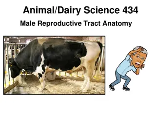
THE MALE REPRODUCTIVE SYSTEM: Composed of: testes, epididymis, vas deferens, seminal vesicles, prostate gland, bulbourethral glands, urethra, and penis.
THE MALE REPRODUCTIVE SYSTEM: 2 Testes: outside abdominal cavity in the scrotum sperm production needs a lower body temp sperm is produced inside tubes called seminiferous tubules produce male sex hormones testosterone, and androgens maturing sperm are moved to the epididymis
THE MALE REPRODUCTIVE SYSTEM: 2 Epididymis: area where sperm mature (coiled tubes) then moved to the ductus (vas) deferens 2 Vas Deferens: sperm stored here (the part close to the epididymis) leads to urethra long tube from epididymis to the urethra
THE MALE REPRODUCTIVE SYSTEM: 2 Seminal vesicles: 2 small glands joins vas deferens at the base of the bladder makes about 60% of the seminal fluid creates a slightly basic (pH 7.5) fluid, high in fructose, amino acids, prostaglandins (hormones that causes contractions of the vagina to help move sperm), and buffers. (buffering keeps the pH constant)
THE MALE REPRODUCTIVE SYSTEM: Prostate: found around urethra contributes alkaline (basic) solution to the semen. 2 Bulbourethral glands (also called cowper s gland): adds to seminal fluid 2 small glands secretes alkaline fluid to neutralize urine in urethra (pre-ejaculate). seminal fluid + sperm = semen
THE MALE REPRODUCTIVE SYSTEM: Urethra: double purpose tube (semen and urine), but never at same time Semen is expelled out of urethra by rhythmic muscular contractions (ejaculation) Penis: becomes erect and firm to allow semen to be deposited in the vagina near the cervix
THE MALE REPRODUCTIVE SYSTEM: The sperm is specially designed for its role. A haploid nucleus Acrosome (contains enzymes that digest the barriers surrounding the egg) Mitochondria for energy production A tail for swimming
THE FEMALE REPRODUCTIVE SYSTEM Composed of: ovaries, fimbria, oviducts (uterine tubes), the uterus, cervix, vagina, clitoris, labia Secondary sexual characteristics are not directly involved in the reproductive system so are not looked at here. 2 Ovaries: Produce the eggs from follicles. Produce estrogen from follicles. Produce progesterone from the corpus luteum.
THE FEMALE REPRODUCTIVE SYSTEM 2 Fimbria: Has cilia to sweep the egg into the oviduct 2 Oviducts (tubes): Contain cilia to push the egg from the ovaries to the uterus. The egg is fertilized while in the oviducts Uterus: Has a thick muscular wall, shaped like an upside down pear. Lined with a layer called the endometrium. The fertilized egg attaches to the wall and develops into a fetus here.
THE FEMALE REPRODUCTIVE SYSTEM Cervix: Produces mucus which depending on the hormones present can either help sperm along or stop them entirely. During pregnancy closes off to hold the baby in. Vagina: Also called the birth canal. Expands in length and width during sex. When stimulated during sex it secretes alkaline mucus to help the sperm survive and to reduce friction with the penis. The concentration of nerve endings are shallow.
THE FEMALE REPRODUCTIVE SYSTEM Clitoris: Sexually sensitive organ located above the vaginal opening. Contains erectile tissue and it becomes enlarged when stimulated. Stimulation can lead help with the female orgasm. Vulva: This is the name given to the external female genitalia.
THE MENSTRUAL CYCLE Is a monthly sequence of ovarian and uterine events controlled by hormones. The hypothalamus in the brain releases the hormone GnRH (gonadotropin-releasing hormone) and this stimulates the anterior pituitary to release FSH and LH. These are two hormones that act on the ovaries: FSH - Follicle Stimulating Hormone - stimulates the follicle to mature and causes it to produce estrogen. LH - Leutinizing Hormone - maintains the corpus luteum and causes it to produce progesterone.
THE MENSTRUAL CYCLE The average cycle lasts 28 days. During the cycle the ovary releases an egg and the uterus is prepared for embryo implantation. By convention we consider the start of the cycle to be the shedding of the endometrium. The cycle consists of two phases: The follicular phase. (days 1-14) Day 1-7: High levels of FSH and low levels of LH FSH stimulates a follicle to grow in the ovary
THE MENSTRUAL CYCLE The endometrium lining of the uterus is shed and exits through the vaginal opening. (menstruation) Developing follicle produces low levels of estrogen and progesterone. Low levels of estrogen keep the LH levels low.
THE MENSTRUAL CYCLE Day 7-14: Menstruation is over and the uterine lining begins to rebuild. As the follicle grows in the ovary it produces more estrogen. Higher estrogen levels increase LH levels and lowers FSH levels. The LH level and FSH level eventually spike causing the egg in the follicle to be released from the ovary (ovulation).
THE MENSTRUAL CYCLE The luteal phase (Day 14-28): Ovulation happens around day 14. The egg is transported down the oviduct. The ruptured follicle develops into the corpus luteum. LH, FSH and estrogen levels drop. The corpus luteum starts producing progesterone in large amounts, and small amounts of estrogen. The progesterone level suppress the FSH and LH levels inhibiting further follicle development.
THE MENSTRUAL CYCLE Rise in progesterone levels converts the thickening uterine lining to an actively secreting tissue with a well-developed blood supply. PREGNANCY: If a fertilized egg implants the corpus luteum remains active for a while keeping progesterone and estrogen levels high. NOT PREGNANT: If fertilization does not occur, then the corpus luteum degenerates and the progesterone and estrogen levels fall causing menstruation to start.
FERTILITY AND FERTILIZATION During copulation, the erect penis ejaculates semen (average 3 or 4 mls of semen containing up to 300 million sperm) into the vagina. The sperm swim through the vagina, uterus, and uterine tubes where fertilization usually takes place. Both sperm and eggs only live for a few days, so fertilization can succeed only if copulation occurs within a couple of days before or after ovulation.
FERTILITY AND FERTILIZATION During fertilization, a few hundred sperm swarm the egg. Enzymes released by the all the sperms acrosomes digest the outer corona radiata and zona pellucida layers of the unfertilized egg. This allows a single sperm cell to penetrate the egg and fertilize it. The two haploid cells fuse into a single diploid cell giving it all of the genes of a new human. Permanent contraception can be achieved by sterilization, either by vasectomy or tubal ligation.
FERTILITY AND FERTILIZATION Most temporary contraception methods prevent ovulation or prevent sperm and egg from meeting. Abortion removes the embryo from the uterus, but is not considered a contraceptive device because it terminates, rather than prevents, pregnancy.
HOW DO HUMANS DEVELOP? A fertilized egg (zygote) develops into a blastocyst and implants in the endometrium. During implantation, the outer blastocyst layer forms the chorion, while the inner cell mass forms the amnion, yolk sac, and embryonic disc. During the third week of development, chorionic villi extend into the endometrium of the uterus.
HOW DO HUMANS DEVELOP? During the fourth week of development, the endoderm forms a tube that will eventually form the digestive tract. The umbilical cord also forms between the embryo and the placenta of the mother. During the sixth week of development, the embryo displays prominent chordate features (notochord, tail, gill grooves), rudimentary eyes, and a developing brain.
HOW DO HUMANS DEVELOP? By the eighth week of development, most of the major organs have developed. The gonads form and develop, producing testosterone or estrogen that will affect future embryonic development. The developing embryo is now called a fetus. Over the next seven months, the fetus continues to grow and develop, with its organs becoming more functional.
HOW DO HUMANS DEVELOP? During pregnancy, the mammary glands within the mother s breasts grow under the influence of estrogen and progesterone. After birth, infant suckling stimulates the release of prolactin and oxytocin, which triggers milk secretion and release from the mammary glands. Nine months of fetal development culminates in labor and delivery, which occurs through the interplay of estrogen, progesterone, steroid hormones produced by the fetus, oxytocin, and uterine stretching.