Respiratory Acidosis and Alkalosis in Clinical Practice
Respiratory acidosis and alkalosis play critical roles in systemic pH regulation. This article delves into the mechanisms, clinical features, and management of these acid-base disturbances. It explores the factors influencing Paco2 levels, consequences of respiratory acidosis, and the significance of arterial pH balance in maintaining homeostasis.

Download Presentation

Please find below an Image/Link to download the presentation.
The content on the website is provided AS IS for your information and personal use only. It may not be sold, licensed, or shared on other websites without obtaining consent from the author.If you encounter any issues during the download, it is possible that the publisher has removed the file from their server.
You are allowed to download the files provided on this website for personal or commercial use, subject to the condition that they are used lawfully. All files are the property of their respective owners.
The content on the website is provided AS IS for your information and personal use only. It may not be sold, licensed, or shared on other websites without obtaining consent from the author.
E N D
Presentation Transcript
RESPIRATORY ACIDOSIS AND ALKALOSIS Dr. BIBYMOL JOSEPH JUNIOR RESIDENT GENERAL MEDICINE
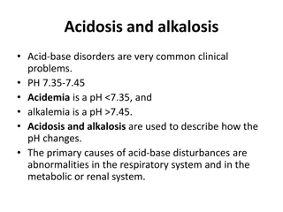
NORMAL ACID BASE HOMEOSTASIS Systemic arterial pH is maintained between 7.35 and 7.45 by extracellular and intracellular chemical buffering together with respiratory and renal regulatory mechanisms. The control of arterial ??2tension (Paco2 ) by the Central Nervous System (CNS) and respiratory system and the control of plasma bicarbonate by the kidneys stabilize the arterial pH by excretion or retention of acid or alkali.
The metabolic and respiratory components that regulate systemic pH are described by the HENDERSON-HASSELBALCH EQUATION pH = 6.1+ log HCO3-/Paco2 x 0.03001 Under most circumstances, CO2 production and excretion are matched, and the usual steady-state Paco2 is maintained at 40 mm Hg. Underexcretion of CO2 produces hypercapnia,and overexcretion causes hypocapnia.
The Paco2 is regulated primarily by neural respiratory factors and is not subject to regulation by the rate of CO2 production. Hypercapnia is usually the result of hypoventilation rather than of increased CO2 production. Increases or decreases in Paco2 represent derangements of neural respiratory control or are due to compensatory changes in response to a primary alteration in plasma HCO3.
RESPIRATORY ACIDOSIS Respiratory acidosis is the acid base disturbance initiated by an increase in Paco2. The level of Paco2 is determined by the rate of CO2 production (VCO2) and the rate of alveolar ventilation (VA) Paco2 = K xVCO2/VA An increase in Paco2 can occur by one of the three possible mechanisms, Presence of excess CO2 in inspired air Decreased alveolar ventilation Increased production of CO2 in the body
CLINICAL FEATURES Clinical features vary according to the severity and duration pf respiratory acidosis, the underlying disease,and whether there is accompanying hypoxemia. A rapid increase in Paco2 may cause anxiety, dyspnoea, confusion, psychosis and hallucinations and may progress to coma. In chronic hypercapnia lesser degrees of dysfunction may occur; sleep disturbances, loss of memory, daytime somnolence, personality changes, impairment of coordination and motor disturbances such as tremor,myoclonic jerks,and asterixis. Headaches, papilledema, abnormal reflexes and focal muscle weakness are due to vasoconstriction secondary to loss of vasodilator effects of CO2.
CAUSES Central Airway Drugs (anesthetics,morphine,sedatives) Obstruction Stroke Asthma Infection
Parenchyma Neuromuscular Emphysema Poliomyelitis Pneumoconiosis Kyphoscoliosis Bronchitis Myasthenia Adult respiratory distress syndrome Muscular dystrophies Barotrauma
Miscellaneous Obesity Hypoventilation Permissive hypercapnia
Depression of the respiratory center by a variety of drugs, injury, or disease can produce respiratory acidosis. This may occur acutely with general anesthetics, sedatives, and head trauma or chronically with sedatives, alcohol, intracranial tumors, and the syndromes of sleep- disordered breathing including the primary alveolar and obesity-hypoventilation syndrome. Abnormalities or disease in the motor neurons, neuromuscular junction, and skeletal muscle can cause hypoventilation via respiratory muscle fatigue.
Mechanical ventilation, when not properly adjusted and supervised, may result in respiratory acidosis, particularly if CO2 production suddenly rises (because of fever, agitation, sepsis, or overfeeding) or alveolar ventilation falls because of worsening pulmonary function. High levels of PEEP in the presence of reduced cardiac output may cause hypercapnia as a result of large increases in alveolar dead space. Permissive hypercapnia may be used to minimize intrinsic positive end-expiratory pressure in acute lung injury/ acute respiratory distress syndrome and severe obstructive lung disease.
Acute hypercapnia follows sudden occlusion of the upper airway or generalized bronchospasm as in severe asthma,anaphylaxis,inhalational burn,or toxin injury. Chronic hypercapnia and respiratory acidosis occur in end-stage obstructive lung disease. Restrictive disorders involving both the chest wall and the lungs can cause respiratory acidosis because the high metabolic cost of respiration causes ventilatory muscle fatigue. Advanced stages of intrapulmonary and extrapulmonary restrictive defects present as chronic respiratory acidosis.
DIAGNOSIS The diagnosis of respiratory acidosis requires the measurement of Paco2 and arterial pH. In acute respiratory acidosis, there is a compensatory elevation (due to cellular buffering mechanisms) in HCO3 , which increases 1 mmol/L for every 10-mmHg increase in Paco2 . In chronic respiratory acidosis (>24 h), renal adaptation increases the [HCO3 ] by 4 mmol/L for every 10-mmHg increase in Paco2 . The serum HCO3 usually does not increase above 38 mmol/L.
A detailed history and physical examination often indicate the cause. Pulmonary function studies including spirometry, diffusion capacity for carbon monoxide, lung volumes,and arterial Paco2 and O2 saturation,usually make it possible to determine if respiratory acidosis is secondary to lung disease. The workup for non-pulmonary causes should include a detailed drug history, measurement of hematocrit, and assessment of upper airway, chest wall, pleura, and neuromuscular function.
TREATMENT The management of respiratory acidosis depends on its severity and rate of onset. Acute respiratory acidosis can be life-threatening, and measures to reverse the underlying cause should be undertaken simultaneously with restoration of adequate alveolar ventilation. This may necessitate tracheal intubation and assisted mechanical ventilation. Oxygen administration should be titrated carefully in patients with severe obstructive pulmonary disease and chronic CO2 retention who are breathing spontaneously.
When oxygen is used injudiciously, these patients may experience progression of the respiratory acidosis causing severe acidemia. Aggressive and rapid correction of hypercapnia should be avoided, because the falling Paco2 may provoke the same complications noted with acute respiratory alkalosis (i.e., cardiac arrhythmias,reduced cerebral perfusion,and seizures).
The Paco2 should be lowered gradually in chronic respiratory acidosis, aiming to restore the Paco2 to baseline levels and to provide sufficient Cl and K+ to enhance the renal excretion of HCO3-. Chronic respiratory acidosis is frequently difficult to correct, but measures aimed at improving lung function should be the primary focus of treatment.
RESPIRATORY ALKALOSIS Acid base disorder initiated by a decrease in Paco2 and increase in the HCO3-/Paco2 ratio ,thus increasing the pH. This occurs when there is excessive loss of CO2 by alveolar hyperventilation. Hypocapnia develops when a sufficiently strong ventilatory stimulus causes CO2 output in the lungs to exceed its metabolic production by tissues.
Plasma pH and HCO3- appear to vary proportionately with Paco2 over a range from 40-15 mmHg Relationship between a) arterial H+ concentration and Paco2 is -0.7 mmol/L per mmHg ( 0.01pH unit/mm Hg) b) plasma HCO3- and Paco2 is 0.2 mmol/L per mmHg
Hypocapnia sustained for > 2-6 hrs is further compensated by decrease in renal ammonium and titratable acid excretion and a reduction in the filtered HCO3- reabsorbtion. Full renal adaptation to respiratory alkalosis may take several days and requires normal volume status and renal function. In chronic respiratory alkalosis a 1 mmHg decrease in Paco2 causes a 0.4-0.5 mmol/L drop in HCO3- and 0.3mmol/L decrease in H+ ( 0.003 increase in pH )
EFFECTS OF RESPIRATORY ALKALOSIS Reduced blood flow as a result of rapid decline in Paco2 may cause dizziness, mental confusion and seizures,even in the absence of hypoxemia. The CVS effects of acute hypocapnia in the conscious human are generally minimal,but in anaesthetized or mechanically ventilated patients cardiac output and blood pressure may fall. Cardiac arrythmias may occur in patients with and heart disease. Acute respiratory alkalosis causes intracellular shifts of K+ and PO4- and reduces free Ca2+ by increasing the protein bound fraction.
Chronic respiratory alkalosis is the most common acid- base disturbance in critically ill patients. Respiratory alkalosis is common during mechanical ventilation.
CAUSES Central nervous system stimulation Hypoxemia and tissue hypoxia Pain High attitude Anxiety, psychosis Pneumonia ,pulmonary edema Fever Aspiration Cerebrovascular accident Severe anaemia Meningitis, encephalitis Tumours
Drugs or hormones Stimulation of chest receptors Pregnancy , Progesterone Hemothorax Salicylates Flail chest Cardiac failure Cardiac failure Pulmonary embolism
Miscellaneous Septicemia Hepatic failure Mechanical ventilation Heat exposure Recovery from metabolic acidosis
The hyperventilation syndrome may be disabling. Paresthesia ,circumoral numbness, chest wall tightness or pain ,dizziness, inability to take up an adequate breath,and rarely tetany. ABG shows an acute or chronic respiratory alkalosis, often with hypocapnia in the range of 15-30 mmHg with no hypoxemia. CNs diseases or injury can produce several patterns of hyperventilation and sustained Paco2 levels of 20-30 mmHg .
Salicylates are the most common cause of drug induced respiratory alkalosis as a result of direct stimulation of medullary chemoreceptor. The methylxanthines, aminophylline and theophylline can stimulate increase the ventilatory response to CO2. ventilation and Progesterone increase ventilation and lowers arterial PCO2 by as much as 5-10 mmHg ,therefore chronic respiratory alkalosis is a common feature of pregnancy.
Respiratory alkalosis is also prominent in liver failure,and severity correlates with degree of hepatic insufficiency. Respiratory alkalosis is often an early finding in gram negative septicaemia, before fever, hypoxemia or hypotension develops.
DIAGNOSIS Depends on arterial pH and Paco2 measurement. The plasma K+ is often reduced and Cl- is increased. In acute respiratory alkalosis ,HCO3- concentration falls by 2 mmol/L for each 10 mmHg decrease in Paco2. In chronic respiratory alkalosis ( >3-5 days) HCO3- by 4-5 mmol/L for each 10 mmHg decrease in Paco2. a decline in Paco2 reduces the serum
Moreover the compensatory reduction in the plasma HCO3- concentration is so effective in chronic respiratory alkalosis that the pH does not decline significantly from normal value. Chronic respiratory alkalosis is the only acid base disorder that may return the pH to the normal value.
TREATMENT Management directed towards the alleviation of underlying disorder. If respiratory alkalosis complicates ventilatory management ,changes in dead space, tidal volume and frequency can minimize the hypocapnia. Patients with hyperventilation syndrome can benefit from reassurance, rebreathing from a paper bag during symptomatic attacks and attention to underlying psychological stress Antidepressants and sedatives are not recommended. Beta blockers may ameliorate peripheral manifestations of hyperadrenergic state.