Respiratory Assessment in Critical Care: Basics and Pathophysiology
Explore the essentials of respiratory assessment in critical care, covering the upper and lower respiratory tracts, conducting airways, respiratory airways, and the importance of air conditioning. Understand the reasons for assessment, from gaining a baseline to diagnosing conditions. Delve into the physiology of respiratory ducts and the intricacies of dead space in the lungs for efficient gas exchange.

Download Presentation

Please find below an Image/Link to download the presentation.
The content on the website is provided AS IS for your information and personal use only. It may not be sold, licensed, or shared on other websites without obtaining consent from the author. If you encounter any issues during the download, it is possible that the publisher has removed the file from their server.
You are allowed to download the files provided on this website for personal or commercial use, subject to the condition that they are used lawfully. All files are the property of their respective owners.
The content on the website is provided AS IS for your information and personal use only. It may not be sold, licensed, or shared on other websites without obtaining consent from the author.
E N D
Presentation Transcript
Respiratory Assessment Basic Assessment in Critical Care
Reasons for Assessment To gain a baseline To recognise changes To diagnose
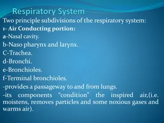
Back to Basics Upper Respiratory Tract: Nose Nasal cavity Paranasal sinuses Nasopharynx Pharynx Oropharynx
Back to Basics Lower Respiratory tract: Larynx Trachea Bronchi Bronchioles Alveoli
Conducting Airways Nasal cavities Pharynx Larynx Trachea Left and Right main Bronchi Lobar Bronchi Segmental Bronchi Bronchioles Terminal Bronchioles
Respiratory Airways Respiratory Bronchioles Alveolar Ducts Alveolar Sacs Alveolar
Air Conditioning Habitually/obligatory nasal breathers for humidification Nasal flare breathing pre- mouth breathing Extensive vascularization of the nasal passage (nose bleed?) Air uptake via nasal passages has initial filter via cilia: Debris remains within the nasal opening Cilia wafts debris to pharynx to be swallowed
Air Conditioning Respiratory mucosa lines the conducting airways Mucous production and cilia work to trap debris and micro- organisms Aim to keep the airways moist If debris pass into the trachea, cilia waft towards pharynx Debris within the bronchioles and alveoli managed by pneumocytes and macrophages
Respiratory Ducts Approximately 150 million alveoli per lung Each surrounded by a network of capillaries and fibres Simple squamous epithelial cells for gaseous exchange Pneumocytes I and II
Pathophysiology Remember dead space!! Anatomical dead space: Consists of upper airways and larger airways unable to make gas exchange. ~150mls in an 70 kg adult or around 2-2.5mls/kg/IBW Alveolar dead space: Alveolar ventilated but not perfused (V/Q mismatch) Physiological dead space: Sum of both of the above, or effects such as pulmonary oedema, excess secretions and aspiration
Pathophysiology Pneumocytes: Type 1: Cover 90-95% of alveolar surface area Squamous cell Type 2: Small percentage of these Involved in surfactant production
Gas exchange Large capillary network surrounding each alveoli Single cell exchange Simple diffusion of both gases from higher concentration to a lower concentration Utilised with the Bohr and Haldane effect
Gas exchange Bohr Effect: Affinity of oxygen binding to haemoglobin Related to temperature and pH of the blood Haldane Effect: Once deoxygenation takes place, the affinity of the blood to uptake carbon dioxide is increased
Methods of Assessment Look: Colour central colour Any cyanosis? Red or flushed face? Patient condition Signs of perspiration? Able to speak in full sentences (if no ETT or tracheostomy)? Restless? Confused? Distressed? Patient s posture?
Methods of Assessment Work of breathing: Respiratory rate? Respiratory pattern: Regular or irregular? Chest movement: Unilateral? Bilateral? Trachea central? Depth of breathing: Shallow breathing? Apical breathing? Bulging neck veins?
Methods of Assessment Use of accessory muscles: Scalane Pectoralis Trapezius Sternocleidomastoid External intercostals
Methods of Assessment Listen: (before auscultation) Any audible sounds? Stridor - narrowing or obstruction of upper airway Wheeze narrowing of lower airways Ruttling secretions present Are there upper airway secretions or obvious issues?
Breathing Patterns Tachypnoea is an abnormally rapid rate of breathing 20 bpm] and is usually one of the first indications of respiratory distress Bradypnoea is an abnormally slow rate of breathing 12 bpm], which can indicate severe deterioration in the patient s condition. Possible causes include fatigue, hypothermia, and central nervous system depression and drugs such as opiates
Breathing Patterns Orthopnoea is a condition in which the person must stand or sit in an upright position to breathe comfortably. It can often occur in many conditions including asthma, pulmonary oedema and emphysema Cheyne-Stokes respiratory pattern periods of apnoea alternate with periods of hyperpnoea. Causes include LVF and cerebral injury, and sometimes seen in patients at the end stages of life
Breathing Patterns Kussmaul Breathing [air hunger] deep rapid respirations due to stimulation of the respiratory centre in the brain caused by metabolic acidosis Biot s respirations rapid deep breathing with abrupt pauses. Severe CNS damage
Methods of Assessment Auscultation: Normal breath sounds: Tracheal/bronchial high pitch, loud, hollow, pause between inspiration and expiration, heard over trachea and large airways Vesicular low pitch, no break between inspiration and expiration, heard over periphery of lung Brochovesicular combination of the two above, heard near the major airways in most other parts of the lung
Methods of Assessment Auscultation of abnormal sounds: Crackles (fine) high pitched rustles relating to the reopening of the small airways or the relating to intra-alveolar fluid Crackles (coarse) low pitch, can be loud, heard over the larger airways, relating to sputum or fluid in these areas Expiratory Wheeze whistling sound heard on expiration, relating to air being pushed through a narrowed airway. Bronchoconstriction effects from asthma, anaphylaxis and toxic gas inhalation Inspiratory wheeze (Stridor) relates to obstruction in major airways such as foreign body, laryngeal oedema, epiglottitis, tumour.
Methods of Assessment Pleural friction a rough, grating and crackling sound heard on both inspiration and expiration. Heard over areas where there is pleural inflammation and friction between the visceral and parietal pleura Increased breath sounds consolidation or relating to fibrosis, pneumonia or atelectasis Decreased breath sounds low airflow from mucous or obstruction, increase chest wall thickness, pleural effusion, hyperinflation
Methods of Assessment Feel: Can you feel any obvious secretions? Palpate the trachea: Is there a mediastinal shift? Feel the expansion of each lung: Is there equal air entry and expansion?
Methods of Assessment Sputum: Mucoid white, clear, relating to Asthma, COPD, viral pneumonia Mucopurulent yellow, relating to chronic bronchitis, acute bacterial infection (if increase in WBC) Purulent yellow or green, associated to bronchiectasis, lung abscess, pneumonia Brown old blood, Klebsiella pneumonia Red present blood, bronchiectasis, TB, lung cancer Haemoptysis coughing blood from traceo-bronchial tree Pink and frothy pulmonary oedema
Methods of Assessment Tools to assess: Pulse Oximetry reasonable non-invasive assessment of peripheral arterial saturation
Methods of Assessment Arterial blood gases accurate but invasive assessment of gaseous exchange and acid-base status of a patient