Respiratory System Malfunctions and Their Impact
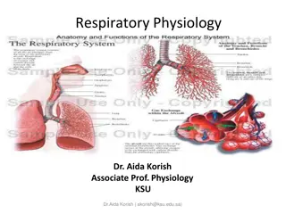
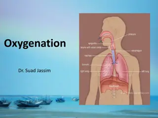
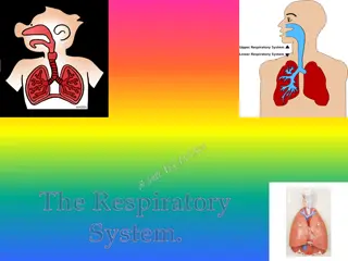
The respiratory system consists of key components such as the larynx, trachea, bronchi, alveoli, diaphragm, and more, all working together to facilitate breathing and gas exchange. Malfunctions in this system can have significant impacts on individuals' health and well-being, affecting oxygen supply, carbon dioxide removal, and overall respiratory function.

Download Presentation

Please find below an Image/Link to download the presentation.
The content on the website is provided AS IS for your information and personal use only. It may not be sold, licensed, or shared on other websites without obtaining consent from the author.If you encounter any issues during the download, it is possible that the publisher has removed the file from their server.
You are allowed to download the files provided on this website for personal or commercial use, subject to the condition that they are used lawfully. All files are the property of their respective owners.
The content on the website is provided AS IS for your information and personal use only. It may not be sold, licensed, or shared on other websites without obtaining consent from the author.
E N D
Presentation Transcript
LO2: The respiratory system, malfunctions and their impact on individuals. Knowledge Organiser Unit 4
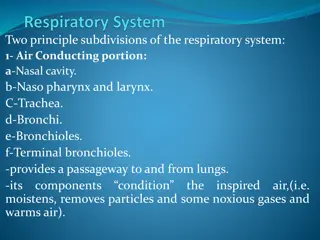
Cartilage: strong, stretchy connective tissue between bones. It isnt as hard and rigid as bone, but less flexible than muscle tissue. Structure of the respiratory system Membrane: thin sheet of body tissue or layer of cells acting as a barrier, lining or partition to separate structures or organs. The respiratory system consists of; Larynx: connects the back of the nose and the trachea , forming an air passage to the lungs. Trachea, bronchi and bronchioles: trachea also known as the windpipe. It starts at the back of the throat (pharynx) and divides into two bronchi, each leading into one of the lungs where they continue to divide to form smaller bronchioles. The trachea and bronchi are tubes that have rings of cartilage along their length to prevent them collapsing open passage for air is maintained. The rings are C shaped in the trachea, with a gap at the back to allow food to travel down the oesophagus. The oesophagus needs to stretch as food passes down. Alveoli: At the end of the tiniest bronchioles are the microscopic alveoli. They are sacs found in clusters at the end of each of the bronchioles. A single sac is called an alveolus. Each alveolus is surrounded by a capillary network where oxygen and carbon dioxide are exchanged through the alveolar membrane. There is an estimated 300 million alveoli in the lungs! Diaphragm: This muscle is anchored to the lower ribs that separates the chest from the abdomen. Intercostal Muscles: these are the muscles found between the ribs. Pleural membranes: Pleural membranes cover the outside of the lungs and line the inside of the chest wall.
Inspiration & Expiration: Structure of Alveoli diagram. The role of pleural membranes: Pleural membranes consist of two layers of thin membrane. They are moist and slippery, having a thin film of liquid between the two layers. It lubricates the surface so that the two pleural layers will slide over each other, this allows the lungs to move easily within the chest cavity. The move with the chest wall as breathing occurs. Role of diagram and intercostal muscles: The function of the respiratory system is to deliver oxygen into the body by breathing in (inspiration) and to remove the waste carbon dioxide by breathing out (expiration). Breathing in air, intercostal muscles pull the ribcage upwards and outwards and the diaphragm flattens inwards. The result of these two movements = increase in volume and a decrease in pressure, this forces air into the lungs so they inflate. Breathing out, the reverse happens diaphragm lifts back into a dome shape, intercostal muscles pull the ribcage inwards and downwards. This process forces carbon dioxide out of the lungs and they deflate. Gaseous exchange: Gaseous exchange is a process that involves the exchange of oxygen and carbon dioxide between capillaries and alveoli.
Diffusion gradients: Diffusion refers to the movement of molecules from an area of high concentration to an area of low concentration. This is the case with gases and for particles dissolved in solutions. Particles diffuse down a concentration gradient, from an area of lower concentration in the red blood cells. Role and structure of alveoli walls The microscopic alveoli are at the end of the tiniest bronchioles and arranged in clusters in the lungs: Exchange of oxygen and carbon dioxide takes place in the alveoli. The capillaries have a lower concentration of oxygen than the alveoli. The walls of the alveoli are very thin (one cell thick), each alveoli is surrounded by capillaries through which gases are exchanged. This results in diffusion of oxygen from an area of higher concentration (the alveoli) to an area of lower concentration in the red blood cells (in capillaries). The structure of alveoli is like a bunch of grapes, which increases their surface area to allow the maximum diffusion of the two gases back and forth making the process very efficient. (see image below) Diffusion allows the oxygen to move out of the alveoli to the capillaries and into the bloodstream and the carbon dioxide to move out of the capillaries into the alveoli and the lungs to be exhaled. 1. Inspiration 2. Exchange of gases lungs to blood 6. Expiration The respiratory process 3. Transport of O & CO in the blood 5. Metabolism 4. Exchanges of gases from blood to tissues
Mitochondria: known as the powerhouses of the cell, they are organelles that act like a digestive system. Taking in nutrients, breaking them down and creating energy rich molecules for the cell. Pyruvate: a molecule that is involved in energy generation . Can be either converted to lactate under anaerobic conditions or broken down into water and carbon dioxide in the presence of oxygen therefore generating large amounts of ATP. Cellular respiration Adenosine triphosphate (commonly known as ATP), is a high energy molecule that is found in every cell. It s job is to store and supply the cell with the energy it needs. Sometimes known as the energy currency of the body. Anaerobic Respiration: This type of respiration takes place if oxygen is not available, it provides less energy. Cellular respiration is a complex set of chemical reactions and processes that take part in the mitochondria to convert biochemical energy from nutrients into ATP and then release waste products. There are two types of respiration that take place in cells to provide energy: Aerobic uses O Anaerobic doesn t need O 2 ATP Glucose pyruvate lactate when oxygen becomes available Above is the equation for Anaerobic respiration. Glycolysis is the process that takes place in the cell cytoplasm breaks down glucose and forms pyruvate, with the production of two molecules of ATP. Pyruvate can be used in either anaerobic respiration if no oxygen is available or in aerobic respiration via a cycle of chemical reactions = more usable energy for the cell. Pyruvic acid supplies energy to cells through the citric acid cycle (also known as Krebs cycle) when oxygen is present (aerobic respiration); when oxygen is lacking, it ferments to produce lactic acid. The lactic acid needs to be oxidised later to carbon dioxide and water to prevent it building up. If oxygen doesn t become available cells die = lactate toxic. Anaerobic respiration is likely to occur when oxygen is in short supply > like when exercising, when muscle cells need lots of energy. Aerobic Respiration: Oxygen and glucose > required for aerobic respiration to take place. It produces waste carbon dioxide and water as well as providing energy. 32 ATP glucose + oxygen carbon dioxide + water Above is the equation for Aerobic respiration. A sugar called glucose, from our food is broken down into water and carbon dioxide and the energy that was holding the glucose molecule together is released.
Respiratory Malfunctions Asthma: Symptoms and effects: Recurring episodes of breathlessness, tightness of chest and wheezing. Asthma attacks episodes of wheezing that require the use of an inhaler to open the airway. Biological explanation: Inflammation of the bronchi that carry air in and out of the lungs, causing the bronchi to be more sensitive than normal. Contact with allergens, something that irritates the lungs known as a trigger (i.e. Cigarette smoke, pollen or dust) it makes airways become narrow, the muscles around them tighten, there is an increase in the production of sticky mucus (phlegm) Cause: The exact cause of asthma is not known and likely to be a combination of factors. It could be genetic, as asthma often runs in families and people who have allergies are higher at risk. Environmental factors also contribute, these include: Exposure to tobacco smoke as a child Triggers like pollen, dust, chemicals like chlorine in swimming pools and air pollution Exposure to smoke in the womb Premature baby (born before 37 weeks gestation or with a low birth weight) Modern hygiene standards we don t build up resistance as environments can be too clean .
Emphysema Emphysema is also known as COPD (chronic obstructive airways disease). Symptoms and effects: Shortness of breath, wheezing Yellow sputum Persistent cough that never seems to go away Frequent chest infections Symptoms get worse over time. Biological Explanation: Airways of lungs become inflamed and narrowed as the air sacs (alveoli) get permanently damaged, it becomes increasingly difficult to breathe out. There is currently no cure for COPD the sooner the condition is diagnosed and appropriate treatment begins, there is a reduced chance of severe lung damage. Cause: Lifestyle choice of smoking is the main cause of COPD and is thought to be responsible for around 90% of cases Some cases of COPD are caused by certain types of fumes, dust, chemical exposure at work = occupational cause Genetic tendency rare, but can be a cause.
Cystic Fibrosis Symptoms and Effects: Lung problems recurring chest infections, persistent inflammation of airways, coughing, wheezing and shortness of breath. Digestive system diarrhoea, diabetes, jaundice and malnutrition as the body struggles to digest and absorb nutrients Could have a serious bowel obstruction in the first few days of life (meconium ileus) this would require surgery to remove the blockage, Biological explanation: Condition is present at birth genetic defect on chromosome 7 that controls movement of salt and water in an out of the cells in the body. The protein produced by the gene causes mucus secreting cells to make really sticky mucus instead of the normal runny mucus. Recurrent infections along with this results in a build up of sticky mucus in the lungs and digestive system. There is no cure over the years the lungs become increasingly damaged and may eventually stop working properly. People with CF have a reduced average life expectancy. Most CF sufferers lead fulfilling lives with successful careers, family life and leisure activities. Cause: Both parents must have a copy of the mutated (faulty) gene. If only one copy of the faulty gene is inherited, the child will be a carrier but not have the condition themselves.
Monitoring treatment and care needs: respiratory malfunctions. There are a variety of methods for testing and monitoring an individual s lung function and to diagnose and assess their condition. Spirometry: a test carried out to measure the breathing capacity of the lungs. It measures the volume of air expired in total and the force of expiration in the first second of breathing out. Used to diagnose COPD and CF. The person has a clip placed on their nose and has to blow into a mouthpiece having inhaled this is repeated at least 3 times over a period of 30-90 minutes. MRI and CT scans: both scans provide high resolution detailed images of the chest and can be repeated over time to monitor changes in the condition. The CT scan is the most sensitive method of detecting emphysema. Peak flow metres: Used to measure the rate of exhalation. For asthma, measurements are taken regularly over time and compared with norms indicating dilation/constriction of airways. MRI Scan: magnetic resonance imaging scan. A strong magnetic field and radio waves used to produce detailed images of the body. CT Scan: computerised tomography scan of the brain, internal organs, blood vessels or bones. Peak flow metre Spirometry
Monitoring treatment and care needs: respiratory malfunctions. Physiotherapy: Used to help restore or improve movement, mobility or function of the body. Consists of exercises, manipulation and massage techniques that can be for specific parts for the body or for movement of the whole body or for lungs and breathing. Used for a wide variety of conditions including COPD and CF. Massage also used to improve quality of life for those with long-term conditions and helps reduce anxiety levels and improve quality of sleep. Inhalers: Preventative inhalers (blue) - used regularly to reduce inflammation and sensitivity of airways. Reliever inhalers (brown/red) muscle relaxants for immediate relief of symptoms. Both types can be used with a spacer device, this gets drugs deeper . Nebuliser may be required if constriction is more severe as this gets the drug deeper into the lungs. Medication: Antibiotics to treat infections; corticosteroids, steroid treatments to relieve symptoms; anti-inflammatory medication reduces swelling and inflammation. Identification of triggers: If asthma is caused by allergens, treatment might involve tests to identify triggers and then de-sensitising injections can be given. Oxygen therapy: (mainly for COPD) Pulmonary rehabilitation = special programme of exercise and education. Ambulatory oxygen therapy use of portable oxygen when walking or other activity. Long-term oxygen therapy use of oxygen at home from a portable oxygen tank. Used with a mask or nasal tubes used for 16 hours a day. Surgery :to remove damaged section of lung or lung transplant only suitable when symptoms are not controlled by medication.
Lifestyle changes and care needs Receiving appropriate treatment and making lifestyle changes can help individuals remain mobile and active by managing their symptoms and minimising the effects of their conditions. Effects on Lifestyle Care Needs Give up smoking Regular check-ups i.e. Asthma clinic Diet well balances to promote maintenance of a healthy weight Vaccinations (CF to avoid infections) Dietary supplements May need to move to a one story house Lack of energy and breathless on any activity Avoid pollution and infections/triggers for asthma Install a stair lift as cannot walk up stairs Having to move around with oxygen cylinder to assist breathing May need to use a wheelchair Emotional and social impacts of not being able to complete daily tasks or go out socially without an oxygen tank, this may lead to depression, anxiety or stress. Oxygen cylinder to assist breathing Home help for daily living tasks. Exam tips: Learn component parts of the respiratory system and be able to explain their function. When answering questions, use the correct terminology. Make sure you know the difference between Aerobic and Anaerobic cellular respiration. Don t mix up causes and effects of malfunctions. Remember the cause is what makes it happen and the effects are the result of it. You must know examples of treatments for each malfunction.