Revelation 22: The River of Life and Tree of Life
In Revelation 22, the vivid imagery of a river of water of life and the tree of life is depicted, symbolizing God's provision of eternal life and healing. The chapter foretells the absence of curse, the reign of God's light, and the eternal service of His servants. Drawing parallels with Genesis, Revelation 22 paints a picture of a restored garden where life's necessities are abundantly provided. Explore the profound significance of these symbols and the promise of everlasting life in God's presence.

Download Presentation

Please find below an Image/Link to download the presentation.
The content on the website is provided AS IS for your information and personal use only. It may not be sold, licensed, or shared on other websites without obtaining consent from the author.If you encounter any issues during the download, it is possible that the publisher has removed the file from their server.
You are allowed to download the files provided on this website for personal or commercial use, subject to the condition that they are used lawfully. All files are the property of their respective owners.
The content on the website is provided AS IS for your information and personal use only. It may not be sold, licensed, or shared on other websites without obtaining consent from the author.
E N D
Presentation Transcript
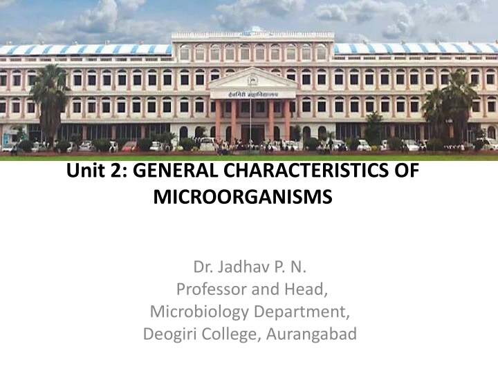
B. Sc. First Year Semester I Paper I Fundamentals of Microbiology Unit 2: GENERAL CHARACTERISTICS OF MICROORGANISMS Dr. Jadhav P. N. Professor and Head, Microbiology Department, Deogiri College, Aurangabad
B. Sc. First Year Semester I Paper I Fundamentals of Microbiology Unit 3: GENERAL CHARACTERISTICS OF MICROORGANISMS Dr. Jadhav P. N. Professor and Head, Microbiology Department, Deogiri College, Aurangabad
Lysogenic cycle The lysogenic cycle (Figure 3), sometimes referred to as temperate or non-virulent infection, does not kill the host cell, instead using it as a refuge where it exists in a dormant state. Following the injection of the phage DNA into the host cell, it integrates itself into the host genome, with the help of phage- encoded integrases, where it is then termed a prophage. The prophage genome is then replicated passively along with the host genome as the host cell divides for as long as it remains there and does not form the proteins required to produce progeny. As the phage genome is generally comparatively small, the bacterial hosts are normally relatively unharmed by this process.
Transition from lysogenic to lytic If a bacterium containing prophage is exposed to stressors, such as UV light, low nutrient conditions, or chemicals like mitomycin C, prophage may spontaneously extract themselves from the host genome and enter the lytic cycle in a process called induction. This process, however, is not perfect and prophage may sometimes leave portions of their DNA behind or take portions of host DNA with them when they re-circularize. If they then infect a new host cell, they may transport bacterial genes from one strain to another in a process called transduction. This is one method by which antibiotic resistance genes, toxin and superantigen-encoding genes and other virulence traits may spread through a bacterial population. Recent work has shown that transition between lytic and lysogenic infection is also dependent on the abundance of phage in an area as they are able to produce and sense small peptides in a process akin to quorum sensing.
LHT System of virus classification The LHT System of Virus Classification is based on chemical and physical characters like nucleic acid (DNA or RNA), Symmetry (Helical or Icosahedral or Complex), presence of envelope, diameter of capsid, number of capsomers. This classification was approved by the Provisional Committee on Nomenclature of Virus (PNVC) of the International Association of Microbiological Societies (1962).
General Characteristics of Fungi Introduction: Fungi is a general term which is used to describe a group of eukaryotes characterised by the absence of chlorophyll and by the presence of rigid cell wall. They can exist either as unicellular in the form of yeast or as multicellular in the form of mould. Edible mushrooms, yeasts, black mold, and the producer of the antibiotic penicillin, Penicillium notatum, are all members of the kingdom Fungi, which belongs to the domain Eukarya.
General Characteristics of Fungi Fungi are eukaryotes possess membrane bound organelles. They can exist both as unicellular and multicellular. They are avascular. Most fungi grow as tubular filament hyphae . A connected mass of hyphae is called mycellium. Protoplasam of hypha (cell) is surrounded by rigid cell wall - The walls of hyphae are reinforced by chitin. The fungal cell wall contains ergosterol rather than cholesterol.
General Characteristics of Fungi Fungi produce a unique form of tubulin in connection with nucelar division. Fungi have a unique biosynthetic pathway for synthesis of lysine. Fungi have a small nuclei with very little repetitive DNA. Mitosis occur without dissolution of nuclear membrane. All are acholorophylous - Fungi are never autotrops because they don t have cholorophyll or choloroplast. May be free living or in intimate relationship - Fungi are usually found as opportunistic saprophytes or in some parasitic or symbiotic relationship with anyother autotrops. Possess characteristic storage range of organic compounds- glycogens, sugar alcohols and lipids.
The fascinating world of fungi- Fungi are Extraordinary organisms which are neither plants, nor animals. One of the most important group of organisms on this planet. Some of the worlds largest and possibly oldest individuals. Hallucinogenic magic mushrooms. Some are silent killers with deadly poisons. A vital ingredient in beer and bread. Decomposers, essential for natural recycling, helping to guarantee life on earth. Miracle cures for disease. Indispensible partners for many plants.
Fungi cell structure and function Fungi are eukaryotes and have a complex cellular organization. As eukaryotes, fungal cells contain a membrane-bound nucleus where the DNA is wrapped around histone proteins. A few types of fungi have structures comparable to bacterial plasmids (loops of DNA). Fungal cells also contain mitochondria and a complex system of internal membranes, including the endoplasmic reticulum and Golgi apparatus. The rigid layers of fungal cell walls contain complex polysaccharides called chitin and glucans. Chitin (fungal cellulose, C22H54N4O21), also found in the exoskeleton of insects, gives structural strength to the cell walls of fungi. The wall protects the cell from desiccation and predators. Most members of the kingdom Fungi are nonmotile.
Fungi Growth The vegetative body of a fungus is a unicellular or multicellular thallus. Dimorphic fungi can change from the unicellular to multicellular state depending on environmental conditions. They display two distinct morphological stages: the vegetative and reproductive. The vegetative stage consists of a tangle of slender thread-like structures called hyphae (singular, hypha ), whereas the reproductive stage can be more conspicuous. The mass of hyphae is a mycelium. It can grow on a surface, in soil or decaying material, in a liquid, or even on living tissue.
Nutrition First, exoenzymes are transported out of the hyphae, where they process nutrients in the environment. Then, the smaller molecules produced by this external digestion are absorbed through the large surface area of the mycelium. As with animal cells, the polysaccharide of storage is glycogen rather than the starch found in plants. Fungi are mostly saprobes (saprophyte is an equivalent term): organisms that derive nutrients from decaying organic matter. They obtain their nutrients from dead or decomposing organic matter, mainly plant material. Fungal exoenzymes are able to break down insoluble polysaccharides, such as the cellulose and lignin of dead wood, into readily-absorbable glucose molecules. Because of their varied metabolic pathways, fungi fulfill an important ecological role and are being investigated as potential tools in bioremediation.
Classification of fungi Martin s (1995) classification of fungi is the most prevalent one. He classified fungi into 3 subdivisions: 1. Schizomycetes (Bacteria) 2. Myxomycetes (Slime moulds) 3. Eumycetes(True fungi)
Yeast Yeast cells are members of the Fungus Kingdom. They are single celled microorganisms (eukaryotic) classified under phyla Ascomycota (sac fungi) and Basidiomyota (higher fungi). Budding - A new yeast cell is formed through mitotic cell division and remains attached as a bud on the old cell until it splits and becomes independent. Here, the parent cell produces an outgrowth that finally splits to become an independent identical cell as the parent cell.
Binary fission In binary fission, no outgrowth (bud) is formed. Rather, through mitosis, the genome replicates and divides followed by the formation of a new plasma membrane and ultimately the cell dividing in to two to form two new cells from the parent cell.
There are useful yeast such as: Baker s Yeast Nutritional Yeast Brewer s Yeast Distiller s and Wine Yeast
Yeast under Microscope Requirements Water Yeast cake Dropper Glass slides and cover slips Stains Yeast cake contains Saccharomyces Cerevisiae (sugar-eating fungus) and can therefore be used to obtain the yeast to observe under the microscope. The following is a procedure that can be used to prepare the specimen for observation. Obtain yeast cake (this can be bought from bakery specialty stores or a supermarket) Cut a small piece of yeast cake and mix with water to form a pasty texture Add a little more water to form a solution Using a dropper, collect and place a drop of the solution on a microscope slide Place a microscope cover slip and observe under high power objective
Observation With Fluorescence microscopy Yeast are among the smallest eukaryotic cells with diameters of between 5 and 10um. For this reason, it is important to view them under high magnification using fluorescence microscopy. Here, 60x or 100x objectives with numerical aperture of 1.4 are recommended for visual observation and maximum brightness and resolution respectively. If recording using a digital camera (e.g. using a 6.8 x 6.8 um sq pixel camera) then a magnification of 60x would be recommended with numerical aperture of 1.4 (Ha ek, 2006).
With Brightfield microscopy When viewing the specimen under high magnification (1000x and above) one will see oval (egg shaped) organism, which are the yeast. It is also possible to observe the buds, which can be seen on some of the yeast cells. If the solution had some sugar, one will also notice some bubbles in the specimen, which are as a result of the fermentation process by the microorganisms.