Role of Normal Flora & Pathogens in GI Infections
Normal flora in the gastrointestinal tract play a crucial role in health and disease. This article delves into the composition of normal flora, their impact on disease prevention, epidemiology of GI infections, and management of pathogens like E. coli, Campylobacter, Yersinia, and Clostridium difficile. It also discusses the microbiological methods for diagnosis of bacterial agents causing diarrheal infections.

Download Presentation

Please find below an Image/Link to download the presentation.
The content on the website is provided AS IS for your information and personal use only. It may not be sold, licensed, or shared on other websites without obtaining consent from the author.If you encounter any issues during the download, it is possible that the publisher has removed the file from their server.
You are allowed to download the files provided on this website for personal or commercial use, subject to the condition that they are used lawfully. All files are the property of their respective owners.
The content on the website is provided AS IS for your information and personal use only. It may not be sold, licensed, or shared on other websites without obtaining consent from the author.
E N D
Presentation Transcript
Prof . Prof .Hanan Hanan Habib & Dr. Microbiology Unit Habib & Dr. Khalifa Microbiology Unit Khalifa Binkhamis Binkhamis
Recall the common normal flora of the GIT Understand the role of the normal flora of the GIT in diseases. Describe the epidemiology, risk factors & host defenses in preventing GI infections. Describe various types of acute diarrheal illnesses, the pathogens that cause them, their clinical presentation, pathogenic mechanism and prevention.
Explain the pathogenesis of E.coli, Campylobacter, Yersinea & Clostridium difficile and their management. Discuss microbiological methods used for the diagnosis of common bacterial agents causing diarrheal infection.
Normal flora are microorganisms that are frequently found in various body sites in normal, healthy individuals. Constituents and number vary according to the age and physiologic status. Able to colonize and multiply under the exiting condition of different body sites. Inhibit competing intruders. Have symbiotic relationship that benefit the host. Can cause disease in immunocompromized patients.
Oral cavity: contains high number of flora which vary from site to site of the mouth. Saliva contain mixed flora :108 organism /ml Stomach : empty stomach has no normal flora in health due to HCL and peptic enzymes Small intestine : very scanty except near colon Colon of adults: 1010 org/gm stool, >90% are Bacteriodes ( anaerobic), 10% other bacteria. Direct effect of diet composition.
Mouth Neisseria spp., Moraxella, Peptostreptococcus. Nasopharynx Neisseria spp., Viridans sterpt. Moraxella, Peptostreptococcus. Stomach Peptosterptococcus, others from mouth. Small intestine Colon Fusobacterium, Eubacterium, Lactobacillus, Enterobacteriaceae, Clostridium, Enterococcus Mouth: Viridans streptococci, Mouth: Candida albicans Nasopharynx: S.pneumoniae, Mouth Nasopharynx N.meningitidis, H.infuenzae, S.pyogenes, S.aureus Stomach Small intestine Colon Pseudomonas, Candida, Clostridium (C. perfringens, C. difficile) Nasopharynx : Stomach: none Small intestine : none Colon: B.fragilis, E.coli, Stomach : Streptococci, Small intestine: scanty, variable Colon: : Bacteriodes, Normal flora ( low virulence) Normal flora ( low virulence) Potential pathogen (carrier) Potential pathogen (carrier)
Many are opportunistic pathogens , eg. perforation of the colon from ruptured diverticulum, feces enter into peritoneal cavity and cause peritonitis Viridans streptococci of oral cavity enters the blood and colonize damaged heart valves. Mouth flora play a role in dental caries. Compromised defense systems increase the opportunity for invasion. Death after lethal dose of radiation due to massive invasion of normal flora.
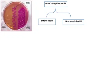
E.coli a facultative flora of colon followed by Klebsiella, Proteus and Enterobacter. Salmonella, Shigella and Yersinia are NOT normal flora of the intestinal tract. Some strains of E.coli ,Salmonella ,Shigella and Yersinia enterocolitica are able to cause diseases in the intestinal tract. E.coli : the most common Enterobacteriacae;
Invasive and inflammatory diarrhea ( Dysentry and /or blood in the stool. Enterotoxin diarrhea with loss of fluid. Some produce systemic illness to multiple organs such as enteric (typhoid) fever. Invasive and cytotoxic cytotoxic strains strains produce Dysentry ) with WBCs Enterotoxin producing strains producing strains cause watery systemic illness due to spread
Acute Diarrheal Illnesses and Food Poisoning Acute Diarrheal Illnesses and Food Poisoning
Acute diarrheal illness is one of the most common problems evaluated by clinicians. A major cause of morbidity and mortality world wide. Most of healthy people have mild illness but other might develop serious squeals so it is important to identify those individuals who require early treatment.
Stool weight in excess of 200 gm/day, or three or more loose or watery stools/day Alteration in normal bowel movement characterized by decreased consistency and increased frequency Less than 14 days in duration.
Viral developed countries Bacterial responsible for most cases of severe diarrhea Protozoan Viral: 70-80% of infectious diarrhea in Bacterial: 10-20% of infectious diarrhea but Protozoan: less than 10%.
1.2 - 1.9 episodes per person annually in the general population 2.4 episodes per child <3 years old annually 5 episodes per year for children <3 years old and in daycare Seasonal peak in the winter.
Infectious Diarrhea: Bacterial infections (eg. Campylobcator, Shigella, Salmonella, Yersinia, Vibrio cholerae & E.coli). Food Poisoning aureus, Clostridium perfringens, Bacillus spp. Traveler Diarrhea E.coli. Antibiotic Associated Diarrhea difficile. Infectious Diarrhea: caused by Viral or Food Poisoning: caused by Staphylococcus Traveler Diarrhea : caused by Enterotoxigenic Antibiotic Associated Diarrhea: Clostridium
Food from restaurant Family member with gastrointestinal symptoms Recent travel to developing countries Patient underlying illness and medication, low stomach acidity, cyst, spores Abnormal peristalsis Low Immunoglobulin A (IgA). Antibiotics decrease the normal flora to less than1012 Median infective dose (ID (ID50 50) )
Enterotoxin mediated Lack of pus in the stool (no gut invasion) No fever Some have rapid onset (<12 hour if due to preformed toxin ingestion) Small intestine affected. Vomiting, non-bloody diarrhea, abdominal cramps. Vibreo cholerae, Staphylococcus aureus, Clostridium perfringens and Bacillus cereus Some viral and parasitic infections. Enterotoxin mediated Small intestine affected.
Invasive Invasive Extension to lymph nodes Incubation period 1-3 days Dysentery syndrome- gross blood and mucous EHEC bloody diarrhea Entameoba histolytica 1-3 wk Pus and blood in the stool Fever due to inflammation Shigella, Salmonella spp., Campylobacter, some E.coli and Entameoba histolytica Affect colonic mucosa
Gram negative curved ( spiral or S-shape ) bacilli . world wide infection ,more common among children Common species : C.jejuni Source meat, person to person transmission can occur. C.jejuni, C. coli, C fetus. Source: dog , cat, birds, poultry ,water, milk,
Incubation period: 2-6 days Lower abdominal pain , watery or dysenteric diarrhea with pus and blood. fever in some patients. Nausea and vomiting are rare Self limiting after 2-6 days. Chronic carrier & outbreaks uncommon. May lead to autoimmune disease like Guillain- Barrie syndrome and extra-intestinal infections eg. reactive arthritis ,bacteremia ,lung infection and others frequently preceded by C.jejuni infection.
Lab diagnosis Use transport media Culture on CAMPY BAP media containing antibiotics. Incubate in microaerophilic atmosphere (5%O210%CO2 85%N ) at 42 C except C.fetus 37 C Identification :Gram stain/culture / biochemical/Serology. Lab diagnosis Treatment: Only severe cases Erythromycin or Ciprofloxacin . .
About 10 -15% of strains of E. coli associated with diarrhea. Other strains associated with extra-intestinal diseases ( septicemia, meningitis & UTI). Based on virulence factors, clinical manifestation, epidemiology and different O and H serotype. Types of Diarrheagenic E. coli : 1. Enterotoxigenic E. coli (E T E C) 2. Enteropathogenic E. coli (E P E C) 3. Enteroinvasive E. coli (E I E C) 4. Enterohaemorrhagic E. coli (E H E C ) 5. Enteroaggregative E.coli (EAEC)
Major cause of Traveler's diarrhea in infant and adult in developing countries due to consumption of contaminated food and water. It has high infective dose Produce heat-labile toxin (LT) and heat-stable toxin (ST) ,each has two fragment (A and B inflammation. LT hyper-secretion of fluid with no cellular injury Symptoms watery diarrhea some time vomiting . Self limiting .No routine diagnostic method required. It has high infective dose 10 106 6- -10 1010 10 A and B) . No invasion or LT leads to accumulation of cAMP, which leads to watery diarrhea, abdominal cramps and
Produce dysentery destruction). Mainly seen in children. Similar to Shigella spp. (non motile, LNF) Transmission :Fecal oral route . Fever, severe abdominal cramp, malaise and watery diarrhea Infective dose 106 dysentery (Penetration, invasion and
Cause infantile diarrhea ( bottle fed infants) Disrupt microvilli and intestinal absorptive function. Outbreak in hospital nurseries and day care centers Low grade fever, malaise, vomiting and watery diarrhea mucous in stool but no blood. watery diarrhea
0157 uremic syndrome (HUS count, hemolytic anemia and kidney failure 0157:H :H7 7 Hemorrhagic diarrhea, colitis and hemolytic HUS) manifested with low Platelet Bloody diarrhea leucocytes Bloody diarrhea, low grade fever and stool with no Fatal disease in young and elderly persons in nursing homes Undercooked hamburgers, unpasteurized dairy products, Apple cider, cookie dough
Cytotoxin : Shiga verotoxin dysenteriae) Cytotoxin : Shiga- -toxin I & II ( verotoxin ) toxin I & II (verotoxin ) (Similar to toxin produced by Shigella verotoxin and and E.coli other than 0157:H7 can cause HUS. Diagnosis by culture on SMAC(sorbitol MacConkey agar), Vertoxin detection by immunological test or nucleic acid testing (NAT). Management Antimicrobial therapy not recommended . Management of HUS required.
Pediatric diarrheal disease Adhering to the surface of the intestinal mucosa. Produce aggregative stacked brick . Produce mucoid, watery diarrhea dehydration and abdominal pain mucoid, watery diarrhea, vomiting, May resolve after two weeks or more .
Mesenteric lymphadenitis in children and septicemia in immunocompromised hosts Common in Europe, USA, Canada .Cat, dog, swine (chitterlings) Survive cold temperatures and associated with transfusion of packed red blood cells. Presented with enteritis, arthritis and erythema nodosum Generalized infection in adult and children 1-5 year, usually mild but in old children and adult mimic appendicitis Growth at 25 -30 C, media: Cefsulodin-Irgasan- Novobiocin (CIN media) Presented with enteritis, arthritis and erythema nodosum
Antibiotic associated diarrhea clindamycin). hospital stay for at least Transmitted from person to person via fecal- oral route Cultured from inanimate hospital surfaces Disruption of the endogenous bacterial flora of the colon Produce toxin A ( effects ) surface epithelial cell receptors leading to inflammation ,mucosal injury and diarrhea. Antibiotic associated diarrhea ( ampicillin, clindamycin). Antibiotic used during the last hospital stay for at least 3 ( ampicillin, cephalosporins 8 weeks ( community acquired) or cephalosporins & & Antibiotic used during the last 8 3 days ( hospital acquired). weeks ( community acquired) or days ( hospital acquired). Cultured from inanimate hospital surfaces. . toxin A ( enterotoxic effects ) and enterotoxic & cytotoxic and B ( cytotoxic ) & cytotoxic B ( cytotoxic ) that can bind to
http://t2.gstatic.com/images?q=tbn:SdD2_7XPEywYzM:http://upload.wikimedia.org/wikipedia/commons/0/0f/Clostridium_difficile_01.pnghttp://t2.gstatic.com/images?q=tbn:SdD2_7XPEywYzM:http://upload.wikimedia.org/wikipedia/commons/0/0f/Clostridium_difficile_01.png http://t2.gstatic.com/images?q=tbn:CrQCk8qf1I0XNM:http://www.netterimages.com/images/vpv/000/000/013/13634-0550x0475.jpg http://t0.gstatic.com/images?q=tbn:pKOwzT7m-DDTuM:http://www.pathconsultddx.com/images/S155986750670818X/gr1-sml.jpg
Patient presents with fever, leukocytosis, abdominal pain and diarrhea Pseudomembrane fibrin, and cellular debris in the colonic mucosa Diagnosis: by enzyme immunoassay (EIA), or nucleic acid testing (NAT). Treatment Metronidazole and supportive treatment Pseudomembrane can result ( fibrin, and cellular debris in the colonic mucosa) and toxic Diagnosis: direct toxin detection from stool can result (neutrophils neutrophils, , ) and toxic megacolon megacolon Treatment Metronidazole oral and supportive treatment oral Vancomycin Vancomycin
Selected Clinical and Epidemiologic Characteristics of Typical Illness Caused By Common Foodborne Pathogens* Pathogen Typical Incubation Period Bacterial Duration Typical Clinical Presentation Assorted Foods Salmonella species 1-3 Days 4-7 Days Undercooked eggs or poultry, produce Undercooked poultry, unpasteurized dairy products Many foods Gastroenteritis Campylobacter jejuni 2-5 Days 2-10 Days Gastroenteritis E. coli, Enterotoxigenic Shigella species 1-3 Days 3-7 Days Gastroenteritis 4-7 Days 1-2 Days Gastroenteritis Produce, egg salad
Listeria monocytogenes Deli meat, hotdogs, unpasteurized dairy products Gastroenteritis, meningitis abortion 2-6 weeks Variable Bacillus cereus1-6 hour Vomiting, Gastroenteritis Fried rice, meats <24 hour Clostridium botulinum 12-72 hour Blurred vision, paralysis Home-canned foods, fermented fish Days-months Staphylococcus aureus 1-6 hour Meats, potato & pork, unpasteurized dairy products. Gastroenteritis, particularly nausea 1-2 Days Yersinia enterocolitica 1-2 Days 1-3 weeks Gastroenteritis, appendicitis-like syndrome Undercooked pork, unpasteurized dairy products.
Stool specimen Microscopy: for the presence of polymorphs or blood may help Culture :on selective media for Salmonella, Shigella & Campylobacter. Culture for Vibreo cholerae, EHEC or Yersinia if suspected. Toxin assay: if C.difficile toxins is suspected. Stool specimen: may help .
Ryan, Kenneth J.. Sherris Medical Microbiology, Seventh Edition. McGraw-Hill Education. Intestinal flora, part of chapter 1 Enteric infections and food poisoning, part of the chapter on Infectious Diseases: Syndromes and Etiologies Clostridium difficile, part of chapter 29 Campylobacter, part of chapter 32 E. coli & Yersinia enterocolitica, part of chapter 33


")


")