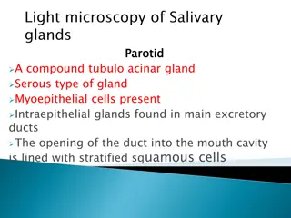
Salivary Gland Diseases and Abnormalities
Explore various salivary gland diseases and developmental abnormalities, including inflammatory lesions, viral diseases, medication-induced dysfunction, and more. Learn about aberrant salivary glands, accessory ducts, and diverticuli, with insights on evaluation and treatment options.

Download Presentation

Please find below an Image/Link to download the presentation.
The content on the website is provided AS IS for your information and personal use only. It may not be sold, licensed, or shared on other websites without obtaining consent from the author. If you encounter any issues during the download, it is possible that the publisher has removed the file from their server.
You are allowed to download the files provided on this website for personal or commercial use, subject to the condition that they are used lawfully. All files are the property of their respective owners.
The content on the website is provided AS IS for your information and personal use only. It may not be sold, licensed, or shared on other websites without obtaining consent from the author.
E N D
Presentation Transcript
Developmental abnormalities Inflammatory and reactive lesions Viral diseases Systemic conditions with salivary gland involvement Medication induced salivary dysfunction Immune conditions Granulomatous conditions Sialorrhea Salivary gland tumors Evaluation of dry mouth Treatment of xerostomia
Developmental abnormalities Absence of glands is rare and occurs in association with other developmental defects, splly malformations of 1stbrachial arch Aplasia xerostomia and dental caries Enamel hypoplasia, congenital absence of teeth and extensive occlusal wear are other symptoms Parotid agensis is reported with several congenital conditions, including hemifacial microstomia, mandibulofacial dysostosis, cleft palate, lacrimoauriculodentodigital syndrome, treacher collins syndrome and anopthalmia Hypoplasia of parotid gland is associated with Melkersson- Rosenthal syndrome
Aberrant salivary glands are salivary tissues that develop at unusual anatomic sites Locations for aberrant glands are middle ear cleft, EAC, neck, posterior mandible, anterior mandible, pituitary and cerebellopontine angle Stafne s cyst: located b/w angle and first molar below the level of inferior alveolar nerve Gland is usually asymptomatic and appears as a round radiolucency Less commonly anterior lingual submandibular salivary glands have been reported Aberrant glands: anterior mandible, appearing as r lucency at tooth apex, extraction sites, b/w roots
Accessory salivary ducts Are common Do not require treatment Frequent location was superior and anterior to normal location of Stenson s duct
Diverticuli It is a pouch or sac protruding from the wall of a duct It leads to pooling of saliva and recurrent sialadenitis Diagnosis: sialography Pts are encouraged to regularly milk the involved gland and promote salivary flow through the duct
Dariers disease Sialography revealed duct dilatation, with periodic stricture affecting the main ducts Symptoms of occasional obstructive sialadenitis are reported
Sialolithiasis Are calcified that form within the secretory system of major salivary glands Factors are inflammation, irregularities in duct system, local irritants and anticholinergic medications may cause pooling of saliva within duct A nidus of salivary organic material becomes calcified and gradually forms a sialolith Submandibular gland is most common site of involvement with 80-90% liths occuring
Common in submandibular gland coz: 1. Torturous corse of Whartons duct 2. Higher calcium and phosphate levels 3. Dependent position of submandibular glands Gout can cause calculi composed of uric acid
Clinical presentation h/o acute, painful and intermittent swelling of affected major gland Degree of symptoms depends on extent of obstruction and presence of secondary infection Eating will initiate swelling Stone totally or partially blocks the flow of saliva, causing saliva pooling within ducts and gland body Since glands are encapsulated, there is little space for expansion and enlargement causes pain
Involved gland is usually enlarged and tender Stasis of saliva may lead to infection, fibrosis and gland atrophy Fistulae, a sinus tract or ulceration may occur over stone in chronic cases Soft tissue examination surrounding duct may show a severe inflammatory reaction Palpation may confirm the presence of a stone Bacterial infections may or may not be superimposed and are more common with chronic obstructions Other complications: acute sialadenitis, ductal stricture and ductal dilatation
r/g examination is needed coz bimanual palpation may not be accessible Poorly calcified liths may not be visible r/g Occlusal view is recommended for submandibular glands parotid gland r/g: AP view with puffed cheek Occlusal film adjacent to duct CT has 10 times the sensitivity of plain film r/g for liths
Calcified phleboliths are stones that lie within a blood vessel Easily mistaken for sialoliths These occur outside the ductal structure Diagnosis: sialography
Treatment Acute phase: supportive Standard care includes, analgesics, hydration, antibiotics and antipyretics In pronounced exacerbations, surgical intervention Liths at or near orifice: removed transorally by milking the gland Deeper stones require surgery Intraglandular stone: removal of the gland Lithotripsy: noninvasive treatment Ultrasonography and extracorporeal lithotripsy to fragment the stone
Mucoceles Is a clinical term that describes swelling caused by accumulation of saliva at site of traumatized or obstructed minor salivary gland duct Classified as extravasation and retention types A large form of mucocele located in floor of mouth is known as ranula
Mucocele Extravasation mucocele: results from trauma to minor glands. Laceration leads to pooling of saliva in adjacent submucosal tissue and consequent swelling Retention mucocele: due to obstruction of minor salivary gland duct by calculus or due to contraction of scar tissue around injured minor salivary gland duct Blockage causes accumulation of saliva and dilation of duct Eventually, an aneurysm like lesion forms, which can be lined by ept of dilated duct
Clinical presentation Extravasation type most frequently occur on lower lip, other regions being buccal mucosa, tongue, FOM,retromolar region Retention cyst: palate or FOM h/o trauma followed by development of lesion Present as discrete painless smooth surfaced swellings ranging from few mm to cm Superficial lesions always have a bluish hue Lesions size vary over time Pts frequently traumatize superficial one, allowing to drain and deflate In these circumstances, they recur
Treatment Surgical excision Removal of associated salivary glands is essential to prevent recurrence Surgical removal may traumatize adjacent glands Intralesional injections of corticosteroids have been successfully used
Ranula Is a large mucocele located on the FOM May be extravasation or retention type Are most commonly associated with sublingual salivary gland duct Etiology: trauma Obstructed salivary gland or ductal aneurysm
Clinical presentation Presents as painless, slow growing, soft and movable mass located in the FOM Lesion forms on one side of lingual frenum If lesion extends deep into soft tissue, it can cross the midline Superficial ranulas can have a blue hue Deep seated one have normal appearing mucosa Large lesion cause deviation of tongue Deep lesion that herniates through the mylohyoid muscle and extends along the facial planes is referred to as plunging ranula and may become large, extending into the neck
Treatment Treated surgically Marsupialization unroofs the lesion, for smaller lesions Recurrent cases, excision of lesion and gland is recommended Intralesional injections of corticosteroids have been successfully used in treatment
Necrotizing sialometaplasia Is a benign self-limiting reactive inflammatory disorder of salivary tissue this lesion mimics a malignancy Initiated by a local ischemic event C/f: has a rapid onset Occur predominantly on palate Lesions initially start as a tender erythematous nodule Once mucosa breaks down, a deep ulceration with yellowish base fprms Even though lesions can be large and deep, pts experience moderate degree of pain
Lesions often occur shortly after oral surgical procedures, restorative dentistry or administration of LA Lesions may also develop weeks after a dental procedure or trauma Treatment: self limiting, lasts approx 6 weeks and heals by secondary intention No specific treatment required Debridement and saline rinses may help healing
Radiation induced pathology Effects of external beam radiation: is standard treatment for head and neck tumors Doses more than or equal to 50 Gy will result in permanent salivary gland damage and symptoms of oral dryness C/p: r therapy is usually delivered in fractionated doses 5 days per week for 6 to 8 weeks Acute effects on salivary function can be recognized within a week of beginning treatment at doses of approx 2 Gy daily and pts will often have c/o oral dryness by end of 2ndweek
Mucositis is a very common consequence of treatment and can be severe to alter radiation therapy If permanent salivary dysfunction develops, pts are at risk of full range of associated oral complications Typically, at doses more than or equal to 50 Gy, dysfunction is severe and permanent Difficulty in speaking, dysphagia and increased caries are common complaints Saliva is minimal and is thick and ropy
Radiation caries: rapidly advancing caries and characteristically occur at incisal or cervical aspect of teeth and wrap around the teeth in an apple core fashion Other complications: candidiasis Sialadenitis Osteonecrosis
Treatment Amifostine, a radioprotective agent Is useful for the preservation of salivary function and for reduction of dry mouth in pts undergoing r therapy Proposed mech involves scavening of free oxygen radicals
Amifostine is dephosphorylated in the circulation by alkaline phosphatase to a pharmacologically active free thiol metabolite Thiol metabolite scavenges free oxygen species generated by radiation Normal tissue is more vascular than tumor and has higher capillary levels of alkaline phosphatase So, the concentration of active thiol metabolite is higher in normal tissue and thus will protect the normal tissue but not cancer Is administered iv 15 to 30 mts prior to each fractionated radiation treatment
Side effects are hypotension, hypocalcemia, nausea and vomitting candidiasis: antifungal agents free of sugar Vaginal clotrimazole troches and dissolved nystatin pwdr Caries: daily prescription strenght topical fluoride is recommended
Effects of internal radiation therapy Disseminated thyroid cancer is treated by removal of thyroid gland To insure all thyroid tissue is removed or destroyed, pts are given radioactive iodine (131I) after surgery R active iodine is taken up not only by thyroid tissue but also by oncocytes in salivary gland tissue R active iodine can cause permanent salivary gland damage and fibrosis resulting in salivary gland hypofunction
Clinical presentation Pts with DTC treated with radioactive iodine may experience xerostomia and decreased salivary gland function Treatment: following administration of r active iodine, pts should suck o lemon drops or chew gum to stimulate salivary flow This will aid in clearing radioactive iodine from salivary glands and potentially decrease gland damage
Allergic sialadenitis Enlargement of salivary glands is associated with exposure to various pharmaceutical agents and allergens Characterisitic feature of allergic reaction is acute salivary gland enlargement and itching over gland Following compounds show gland enlargement: Phenobarbital, phenothiazine, ethambutol, sulfisoxazole, iodine compounds, isoproterenol and heavy metals Allergic sialadenitis is self limiting: avoid allergen, hydration and monitor for secondary infection
Viral diseases : mumps Syn: epidemic parotitis Is caused by a RNA paramyxovirus Transmitted by direct contact with salivary droplets C/p: occurs in children b/w ages of 4 and 6 yrs Incubation period is 2 to 3 weeks Followed by salivary gland inflammation and enlargement, preauricular pain, fever, malaise, headache and myalgia Majority involve parotid glands Skin over gland is oedematous
Salivary gland enlargement is sudden and painful to palapation Ducts are inflammed but without purulent discharge If partial duct obstruction occurs, pt may experience pain when eating One gland can become symptomatic 24 to 48 hrs before another gland does so Swelling is usually bilateral and lasts approx 7 days
Lab: demo of antibodies to mumps S and V antigens and to hemagglutination antigen Serum amylase levels may be elevated Complications: mild meningitis and encephalitis Deafness, myocarditis, thyroiditis, pancreatitis and oophoritis occur less frequently Males may experience epididymitis and orchitis, resulting in testicular atrophy and infertility, if occur in adolesence Treatment: symptomatic
Cytomegalovirus infection Is a beta herpes virus infecting only humans May remain latent after initial exposure and infection c/p: occur In young adult population and presents as an acute febrile illness that causes salivary gland enlargement Transplacental transmission can cause prematurity, low birth weight and congenital malformations Infected newborns and young children suffer from hepatitis, myocarditis, hematologic abnormalities, pneumonitis and nervous system damage Infection is fatal
Those children who survive frequently experience permanent nerve damage resulting in mental retardation and seizure disorders In adults, could be due to reactivation of virus in immunocompromised pts or pts with hematologic abnormalities or HIV infection Advent of highly active antiretroviral therapy for treating HIV infection has resulted in a decline of CMV end organ damage
CMV infected tissue contains large atypical cells with inclusion bodies These cells can be 2 times the normal size and have eccentrically places nuclei, resulting in owl-like appearance Lab: culture, antigen detection, CMV DNA detection Diagnosis of primary infection: combination of IgM anti CMV antibody seropostivity, Ig G seroconversions, and viral culture
Treatment Immunocompetent pts are treated symptomatically Immunocompromised pts require aggressive management Iv gancyclovir, foscarnet or cidofovir
HIV infection Neoplastic and non neoplastic salivary gland enlargement occur with increased frequency in HIV infected pts HIV-salivary gland disease describes xerostomia and benign salivary gland enlargement HIV-SGD is associated with a cluster designation 8 cell lymphocytosis of salivary glands and with diffuse infilterative lymphocytosis sysdrome (DILS)