Salivary Gland Diseases and Disorders
The salivary glands play a crucial role in oral health and can be affected by various developmental, inflammatory, obstructive, and tumorous conditions. Explore the classification, causes, symptoms, and treatments of salivary gland disorders such as xerostomia, sialolithiasis, and more.

Download Presentation

Please find below an Image/Link to download the presentation.
The content on the website is provided AS IS for your information and personal use only. It may not be sold, licensed, or shared on other websites without obtaining consent from the author.If you encounter any issues during the download, it is possible that the publisher has removed the file from their server.
You are allowed to download the files provided on this website for personal or commercial use, subject to the condition that they are used lawfully. All files are the property of their respective owners.
The content on the website is provided AS IS for your information and personal use only. It may not be sold, licensed, or shared on other websites without obtaining consent from the author.
E N D
Presentation Transcript
Dr Gopal Nagargoje OMFS
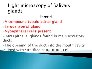
Introduction The salivary glands may be classified as major and minor glands. Major glands are paired glands. They are: (i) Parotid (ii) Sub-mandibularand (iii) Sub- lingual glands. Based on the type of secretion, the salivary glands may be grouped as: (i) Serous, (ii) Mucous and (iii) Mixed.
CLASSIFICATION I. Developmental : 1. Aplasia V. Non-inflammatory : Enlargement of the gland 1. Autoimmune: Sj gren s syndrome and Mickulicz s disease 2. Diabetes mellitus , HIV 3. Nutritional deficiency 2. Atresia 3. Aberrancy II. Salivary gland dysfunction: 1. Xerostomia 2. Sialorrhea III. Obstructive diseases: 1. Sialolithiasis IV. Tumoursof salivary glands : A. Benign tumours: Pleomorphicadenoma Warthin s tumour Myoepithelioma Ductal papilloma B. Malignant tumours : III. Cysts: 1. Extravasationcysts 2. Retention cysts 3. Ranula Basal cell adenoma Canalicularadenoma Iv. Inflammatory Enlargement of the gland 1. Viral: Mumps, coxsackie A, CMV, echovirus, influenza virus 2. Bacterial 3. Allergic Mucoepidermoid carcinoma Adenoid cystic carcinoma Malignant pleomorphicadenoma
Developmental diseases of Salivary Gland 1. Aplasia absence of the gland 2. Atresia absence of the duct 3. Aberrancy ectopic gland
II. Salivary gland dysfunction: Sialorrhoeaor Ptyalism Excessive salivation Minor sialorrhoea can be seen due to local irritation eg. aphthous ulcers or ill fitting Profuse salivation is seen in rabies, heavy metal poisoning or after certain medications like lithium and cholinergic agonists. Dentures, in cerebral palsy 1. Treatment- Anticholinergic medication (atropine) Suggested Surgical Treatment 1. Submandibulargland resection. 2.Transposition of parotid duct. 3. Parotid duct ligation.
Xerostomia This is a subjective sensation of a dry mouth It affects women more than the men (Older) Causes- Antihistamines, decongestants, antidepressants, antipsychotics, antihypertensives, anticholinergicsare known to cause xerostomia. Other causes of xerostomiaare salivary gland aplasia, ageing, excessive smoking, mouth breathing, local radiation therapy, Sj gren s syndrome, HIV infection, etc. Clinically, dry mouth with foamy, thick, ropy saliva can be noticed. The tongue may have leathery appearance and fissures with atrophy of the filiform papillae. These patients are more prone for oral candidiasis, rampant caries Treatment- maintainanceof oral hygiene, use of sialagogues (pilocarpine)
Obstructive diseases: Sialolithiasis Sialolithiasis is the formation of sialolith (salivary calculi, salivary stone) in the salivary duct or the gland resulting in the obstruction of the salivary flow. about 90% of the sialoliths form in the submandibulargland because the long, curved Wharton s duct , the secretion of this gland is higher in calcium content , the position of the gland The sialolith is yellowish white in color, single or multiple, may be round, ovoid or elongated having the size of 2 cms or more in diameter. Contents- hydroxyapatite, octacalcium phosphate, P, ca, bacteria, epithelial cells
Clinical feature- pain and swelling during and after eating the food. Management- The smaller sialoliths, which are located peripherally near the ductal opening may be removed by manipulation. Multiple stones or stones in the gland require the removal of the gland Piezoelectric shockwave lithotripsy to fragment the salivary stones.
CYSTS OF SALIVARY GLAND The cysts of the salivary glands are known as mucoceles. Mucocele It is a swelling due to the accumulation of saliva, as a result of obstruction or trauma to the salivary gland ducts. Types : 1.Extravasation type 2. Retention type. These two types differ in the pathogenesis. The extravasation type is the common one.
Pathogenesis of Mucocele Extravasation cyst Mucous retention cyst Sialolith Trauma (By bitting lip or by Forceps) Obstruction of duct of gland Minor Salivary gland Accumulation of saliva in duct Extravasation of Mucin in connective tissue Dilatation of duct Inflammation (PMN s & Macrophages) Mucocele Granulation Tissue (Wall of Cyst) Mucocele No epithelial lining Epithelial lining present
Clinical features Mucoceles occur as painless swellings, which may be recurrent. (Some patients give the history of increase in size during mealtime). The common sites of occurrences of mucoceles, the extravasation type, are the lower lip and tongue. The retention type of mucocele occurs in palate, cheek, floor of the mouth and maxillary sinus.
The extravasation type occurs in 2nd decade and also in children. The retention type occurs in the middle aged persons. The superficial mucoceles are the bluish translucent seen. They are fluctuant. Usually these cysts are 4 mm to 1 cm in size. The wall is very thin and they rupture easily. The deep mucoceles are firm, well-circumscribed lesions and are present for longer period
TREATMENT Surgical excision is the primary treatment, particularly to prevent recurrence
TREATMENT Aspiration of the fluid from the mucocele will not provide long-termbenefit Intralesional injections of corticosteroids have been used successfully to treat mucoceles.
RANULA It is a special type of mucocele, which occurs in the floor of the mouth. Since the lesion appears like the belly of a frog, it is called Ranula . (Rana-frog) Ranula is formed because of the trauma to sub-mandibular or sub-lingual ducts This develops as a result of mucous extravasation.
It starts as a painless swelling on one side of the floor of the mouth. The swelling is fluctuant, and non-pitting on pressure.
Superficial Ranulas can have a typical bluish hue Deeply seated lesion - normal mucosa Deeply seated Ranula Superficial Ranula
A rare variety, which herniates through the mylohyoid muscle resulting as the swelling of the neck is called plunging ranula . This develops as a result of mucous extravasation.
TREATMENT Treatment of choice - surgical intervention Marsupialization - unroofs the lesion is the initial treatment, especially for smaller lesions. Postsurgicalcomplications Recurrence sensory deficits of the tongue Damage to Wharton s duct
TREATMENT Frequency of recurrence is related to the surgical technique selected Marsupialization: 67% Ranula excision:58% Sublingual gland excision:1% Due to this reason excision of the lesion and the gland should be considered Intralesional injections of corticosteroids have been used successfully in the treatment of ranulas
Inflammatory Enlargement of the gland (Sialadenitis) Inflammation of the salivary glands is known as sialadenitis. Types- Viral, Bacterial, Allergic Viral Infections Mumps (epidemic parotitis) is the most common viral infection affecting the salivary glands. Which is caused by a paramyxo virus. It is an acute, contagious disease, usually affecting the parotid gland. Occasionally, the submandibular or the sublingual glands may also be involved.
This disease is self-limiting one and not dangerous. In these days, the number of cases are reduced, because of the use of the Mumps vaccine. It is a disease of the childhood, but when it affects the adults, it leads to greater complications. This disease has the incubation period of 2-3 weeks.
Clinical features Initially, the patient may suffer from mild fever, headache, chills, vomiting, etc. followed by pain below the ear and sudden onset of firm, rubbery or elastic swelling of the salivary gland. In viral parotitis, the glands of both the sides enlarge
There is no suppuration and no pus discharge from Stenson s duct upon pressure on the gland; however, the papilla may be puffy and red. There is excruciating ear pain during mastication. Xerostomia, trismus, cervical lymphadenitis, tender glands, oedema of the overlying skin may also be present.
Management It is self-limiting. Symptomatic relief can be given by antipyretics. Antibiotics can be given to prevent the secondary infection.
IV. Tumours of salivary glands : A. Benign tumours: Pleomorphic adenoma Warthin s tumour Basal cell adenoma Myoepithelioma Canalicular adenoma Ductal papilloma
Pleomorphic Adenoma Mixed tumor A benign neoplasm consisting of cells exhibiting the ability to differentiate to epithelial cell (ductal & non ductal) and mesenchymal cell (chondroid, myxoid & osseous) Pleomorphic adenoma constitutes more than 50% of all tumours and 90% of all the benign tumours of the salivary glands
Clinical features Parotid gland lower pole, superficially90% Minor Salivary gland8% - mostly palatal F:M = 6:4 4th-6th decade of life 43 yrs mean age Also found in children
Clinical presentation Small, painless, quiscent nodule slowly increases in size, sometimes showing intermittent growth Doesn t fixes todeeper tissue mobile on palpation Skin never ulcerates, although size may reach a fantastic size and of several kilograms
Clinical presentation Pleomorphic adenomaof minor salivary gland of size >1-2 cm creates problem in mastication, talking and breathing Palatal Pleomorphic adenoma may appear fixed to underlying bone but invasive
Treatment Pleomorphic adenomas are treated by surgical excision, excision of the gland with the tumour is performed. Irradiation is contraindicated as the tumour is radioresistant.
Diagnostic Imaging (1) Conventional radiography, (2) Sialography, (3) Ultrasonography, (4) Computerized tomography, (5) Radionuclide imaging, and (6) Magnetic resonance imaging (MRI).
SIALOGRAPHY It is a specialized radiographic procedure performed for detection of disorders of the major salivary glands (usually parotid and submandibular glands. The technique was first performed by Carpy in 1902, using mercury as the contrast agent The technique is employed for examination of both parenchymal (acinar) and ductal abnormalities It involves cannulation and filling with a radio-opaque/contrast agent to make them visible on a radiograph
Indications a. Calculi or foreign bodies b. Determination of the extent of destruction of salivary gland tissue c. Detection of fistulae, diverticuli and strictures d. Detection and diagnosis of recurrent swelling and inflammatory processes e. Demonstration of tumour; its size, location and origin f. Selection of the site for biopsy g. Outline the plane of facial nerve as a guide in planning a biopsy or a dissection
Contraindications Patients with a known allergy or hypersensitivity to contrast media Acute inflammation of the salivary glands Patients scheduled for thyroid function tests in the immediate future.
Contrast Media Lipid soluble or oil-based agents: These agents contain 37% iodine e.g. Ethiadol. Water soluble or water-based agents These agents contain 28 to 38% iodine; e.g. Hypaque 50%, Hypaque M 75%, Renografin 60, 76% Sinografin, Isopaque, Triosil, and Dionosil
Technique Identification of the location of duct orifices Exploration of the duct with a lacrimal probe Cannulation of the ducts: Introduction of the radiographic dye: 0.76-1.0 ml for parotid gland, and 0.5-0.75 ml for submandibular gland. Radiographic projections- Lateral View or Anteroposterior view
(1) Sialogram of normal parotid gland. (2) Sialogram of normal submandibular gland. Note the normal ductal branching pattern in both the sialograms
Observations salivary calculi, stricture or other obstructions appear as radiolucent lesions. In Sj gren ssyndrome, Cherry - blossom or branchless fruit laiden tree appearance Tumours: hand holding the ball appearance.


 Sialogram of normal parotid gland. (2) Sialogram of")