Sheep Brain Dissection Observations: External Structure Analysis
This comprehensive guide provides a detailed examination of the external structures of a sheep brain through dissection. Learn to identify and describe key components such as lobes, meninges, and cortical regions. Explore questions related to motor control, sensory processing, brain connectivity, and more. Engage in hands-on activities and visualizations to enhance your understanding of brain anatomy.

Download Presentation

Please find below an Image/Link to download the presentation.
The content on the website is provided AS IS for your information and personal use only. It may not be sold, licensed, or shared on other websites without obtaining consent from the author.If you encounter any issues during the download, it is possible that the publisher has removed the file from their server.
You are allowed to download the files provided on this website for personal or commercial use, subject to the condition that they are used lawfully. All files are the property of their respective owners.
The content on the website is provided AS IS for your information and personal use only. It may not be sold, licensed, or shared on other websites without obtaining consent from the author.
E N D
Presentation Transcript
Sheep Brain Dissection Observations Please answer in red Group Members:
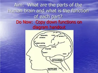
Below is a list of structures you may be asked to identify on an exam, be prepared to describe the functions of all structures Cerebellum Cerebral cortex Frontal cortex Parietal cortex Temporal cortex Occipital cortex Corpus callosum Hypothalamus Medulla oblongata Cranial nerves (general) Meninges Dura mater Arachnoid Pia mater Optic chiasm Pons Spinal cord Thalamus Pituitary
Part 1: External Observations RECORDER: READER: HANDS ON:
External observations Length: Weight: Calculate the size of the donor animal: Describe: Dura mater: Arachnoid (not seen): Pia mater:
Record the approximate sizes of the lobes (estimate first volume and then determine % - using total volume as your denominator) how much of the cortex is made up of each lobe Occipital: Temporal: Frontal: Parietal:
Take a Picture of the Brain from the dorsal view (dura mater removed) and insert below, label all items listed below: Temporal lobe Spinal cord Occipital lobe Frontal lobe Cerebellum Parietal lobe
Respond to the following questions: 1. What does the motor cortex control? 2. What type of information is processed by the sensory cortex? 3. How deep is the longitudinal fissure between the 2 hemispheres? 4. What structure connects these 2 hemispheres of the brain and allows information to be processed by structures in both hemispheres? 5. Describe the appearance of the cerebellum. 6. What is the function of the cerebellum? 7. Describe the appearance of the midbrain structures and the pineal body. 8. What is the function of the pineal body?
Part 2: Ventral Structures RECORDER: READER: HANDS ON:
Ventral Structures: Take a picture of the brain from the ventral view. Insert below and label all items listed below: Frontal lobe Pons Cerebellum Occipital lobe Spinal cord Temporal lobe Optic chiasm Olfactory bulb Pituitary gland Medulla
Ventral Surface Questions 11. Describe the appearance of the olfactory bulbs and include the length of these structures. 12. How does the relative size of these olfactory bulbs compare with those on the human models? 13. Is the sense of smell more important as a protective and a food-getting sense in sheep or in humans? 14. Describe the appearance of the optic chiasm. 15. Explain why signals seen with one eye are processed by the lobe of the opposite side of the brain. 16. What is the function of the pituitary gland? 17. Can you distinguish between the end of the medulla and the beginning of the spinal cord?
Part 3: Cranial Nerves RECORDER: READER: HANDS ON:
Cranial Nerves 18.Describe the appearance of nerve fibers originating on the ventral surface of the brain. 19.How many sets of nerves are you able to identify on your specimen? 20.List the nerves you were able to identify. 21.Why is it so hard to preserve these structures?
Part 4: Internal Structures RECORDER: READER: HANDS ON:
Take a picture of your bisected sheep brain. Label the items listed below. Pituitary gland Frontal lobe Spinal cord Hypothalamus Pons Medulla Occipital lobe Parietal lobe Corpus callosum Grey matter Cerebellum White matter Thalamus
Questions: Internal Structures 22. Describe the appearance of the corpus callosum. 23. What is the function of the corpus callosum? 24. Describe how white and gray matter are arranged in the bisected brain. 25. What is the white matter? 26. What is the gray matter? 27. Describe the appearance of the choroid plexus within the ventricle. What is the function of this structure? 28. What is the function of the cerebrospinal fluid? 29. What is the role of the thalamus? 30. What functions does the hypothalamus regulate? 31. Describe appearance of the cerebellum and the distribution of white and gray matter in the cerebellum. 32. What functions are located in the cerebellum?
Part 5: Coronal Section RECORDER: READER: HANDS ON:
Take a picture of the Coronal (Frontal) Section and label the items listed below Grey matter White matter Thalamus Cerebral cortex Corpus callosum
Clean up Dispose of brain in the plastic bag provided. Return all equipment clean and dried to the back demo table. Wipe the table with a Clorox wipe.


 Section")