Learn about the state of shock, its definition, causes, risk factors, and prevention methods. Shock results from insufficient blood flow to body tissues and can have various types like septic shock, anaphylactic shock, cardiogenic shock, and hypovolemic shock. Recognizing and treating shock promptly is crucial to prevent irreversible organ dysfunction.
Please find below an Image/Link to download the presentation.
The content on the website is provided AS IS for your information and personal use only. It may not be sold, licensed, or shared on other websites without obtaining consent from the author. If you encounter any issues during the download, it is possible that the publisher has removed the file from their server.
You are allowed to download the files provided on this website for personal or commercial use, subject to the condition that they are used lawfully. All files are the property of their respective owners.
The content on the website is provided AS IS for your information and personal use only. It may not be sold, licensed, or shared on other websites without obtaining consent from the author.
E N D
Presentation Transcript
Presenter DeviNanjappan Principal [FACULTY OF NURSING]
Definition Definition Shock is the state of insufficient blood flow to the tissues of the body as a result of problems with the circulatory system. Shock is defined as a state of cellular and tissue hypoxia due to either reduced oxygen delivery, increased oxygen consumption, inadequate oxygen utilization, or a combination of these processes
Incidence Incidence This most commonly occurs when there is circulatory failure manifested as hypotension (ie, reduced tissue perfusion); It is crucial to recognize that a patient in shock can present hypertensive, normotensive, or hypotensive. Shock is initially reversible, It must be recognized and treated immediately to prevent progression to irreversible organ dysfunction.
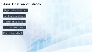
Causes Causes Shock is a common end point of many medical conditions. Shock itself is a life-threatening condition as a result of compromised body circulation Type Cause Low volume Fluid loss such as bleeding or diarrhea Heart Ineffective pumping due to heart damage Obstructive Blood flow to or from the heart is blocked Distributive Due to abnormal flow within the small blood vessels
Risk factors Risk factors Advancing age Have a history of heart failure or heart attack. Have blockages (coronary artery disease) in several of your heart's main arteries. Have diabetes or high blood pressure. female.
Prevention Prevention Shock can be prevented by reducing the incidence of the causes, such as heart failure, injuries, dehydration
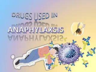
Types of shock Types of shock Septic shock results from bacteria multiplying in the blood and releasing toxins. Common causes of this are pneumonia, urinary tract infections, skin infections (cellulitis), intra-abdominal infections (ruptured appendix), and meningitis. Anaphylactic shock is a type of severe hypersensitivity or allergic reaction. Causes include allergy to insect stings, medicines, or foods (nuts, berries, seafood). Cardiogenic shock happens when the heart is damaged and unable to supply sufficient blood to the body. This can be the end result of a heart attack or congestive heart failure. Hypovolemic shock is caused by severe blood and fluid loss, such as from traumatic bodily injury, which makes the heart unable to pump enough blood to the body, or severe anemia where there is not enough blood to carry oxygen through the body. Neurogenic shock is caused by spinal cord injury, usually as a result of a traumatic accident or injury.
Clinical S/S Clinical S/S Signs common to most forms of shock Pallor, mottled skin, cold extremities, sweating and thirst. Rapid and weak pulse often only detected on major arteries (femoral or carotid). Low blood pressure (BP), narrow pulse pressure, BP sometimes undetectable. Capillary refill time (CRT) > 3 seconds. Cyanosis, dyspnoea, tachypnoea are often present in varying degrees depending on the mechanism. Consciousness usually maintained, but anxiety, confusion, agitation or apathy are common. Oliguria or anuria.
Clinical S/S Clinical S/S Anaphylactic shock Significant and sudden drop in BP Tachycardia Frequent cutaneous signs: rash, urticaria, angioedema Respiratory signs: dyspnoea, bronchospasm Septic shock High fever or hypothermia (< 36 C), rigors, confusion BP may be initially maintained, but rapidly
Clinical S/S Clinical S/S Cardiogenic shock Respiratory signs of left ventricular failure (acute pulmonary edema) are dominant: tachypnoea, crepitations on auscultation. Signs of right ventricular failure: raised jugular venous pressure, hepatojugular reflux,
Diagnosis Diagnosis The aetiological diagnosis is oriented by: The context: trauma, insect bite, ongoing medical treatment, etc. The clinical examination: fever skin pinch consistent with dehydration thoracic pain from a myocardial infarction or pulmonary embolus abdominal pain or rigidity of the abdominal wall from peritonitis, abdominal distension from intestinal obstruction blood in stools, vomiting blood in intestinal haemorrhage subcutaneous crepitations, likely anaerobic infection
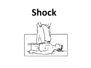
Treatment Treatment In all cases Emergency: immediate attention to the patient. Warm the patient, lay him flat, elevate legs (except in respiratory distress, acute pulmonary edema). Insert a peripheral IV line using a large caliber catheter (16G in adults). If no IV access, use intraosseous route. Oxygen therapy, assisted ventilation in the event of respiratory distress. Assisted ventilation and external cardiac compression in the event of cardiac arrest. Intensive monitoring: consciousness, heart rate, BP, CRT, respiratory rate, hourly urinary output (insert a urinary catheter) and skin mottling.
Treatment Treatment Management according to the cause Haemorrhage Control bleeding (compression, tourniquet, surgical haemostasis) Determine blood group. Priority: restore vascular volume as quickly as possible: Insert 2 peripheral IV lines (2 catheters 16G in adults). Ringer lactate or 0.9% sodium chloride: replace 3 times the estimated losses and/or plasma substitute: replace 1.5 times the estimated losses Transfuse: classically once estimated blood loss represents approximately 30 to 40% of blood volume (25% in children). The blood must be tested (HIV, hepatitis B and C, syphilis, malaria in endemic areas, etc.)
Treatment Treatment Severe acute dehydration due to bacterial/viral gastroenteritis Urgently restore circulating volume using IV therapy with Ringer lactate: As soon as the patient is able to drink (often within 2 hours), provide oral rehydration solution (ORS) as the patient tolerates. Closely monitor the patient;
Treatment Treatment Severe anaphylactic reaction Administer epinephrine (adrenaline) IM, into the anterolateral part of the thigh, in the event of hypotension, pharyngolaryngeal oedema, or breathing difficulties: At the same time, administer rapidly Ringer lactate or 0.9% sodium chloride: 1 litre in adults (maximum rate); 20 ml/kg in children, to be repeated if necessary. repeat IM epinephrine every 5 to 15 minutes. In patients with bronchospasm, epinephrine is usually effective. If the spasm persists give 10 puffs of inhaled salbutamol.
Treatment Treatment Septic shock Vascular fluid replacement with Ringer lactate or 0.9% sodium chloride or plasma substitute. Use of vasoconstrictors: dopamine IV at a constant rate by syringe pump: 10 to 20 micrograms/kg/minute or, if not available epinephrine IV at a constant rate by syringe pump: Use a diluted solution, i.e. add 1 mg epinephrine (1:1000) to 9 ml of 0.9% sodium chloride to obtain a 0.1 mg/ml solution (1:10 000). Start with 0.1 microgram/kg/minute. Increase the dose progressively until a clinical improvement is seen.
Treatment Treatment The objective is to restore efficient cardiac output. The treatment of cardiogenic shock depends on its mechanism. In the event of worsening signs with vascular collapse, use a strong cardiotonic: dopamine IV at a constant rate by syringe pump : 3 to 10 micrograms/kg/minute Once the hemodynamic situation allows (normal BP, reduction in the signs of peripheral circulatory failure), nitrates or morphine may be cautiously introduced. Digoxin should no longer be used for cardiogenic shock, except in the rare cases when a supraventricular tachycardia has been diagnosed by ECG. Correct hypoxia before using digoxin. digoxin slow IV: Adults: one injection of 0.25 to 0.5 mg, then 0.25 mg 3 or 4 times per 24 hours if necessary Cardiac tamponade: restricted cardiac filling as a result of haemopericardium or pericarditis. Requires immediate pericardial tap after restoration of circulating volume. Tension pneumothorax: drainage of the pneumothorax. Symptomatic pulmonary embolism: treat with an anticoagulant in a hospital setting.
Know the differences Know the differences heart attack Blood supply to the heart muscle is reduced or blocked, but the heart keeps beating; however, there may be damage to the heart muscle. Usually, the person knows something is happening, and can talk about his or her symptoms sudden cardiac arrest The electrical system of the heart goes wrong (think of the way the lights flash before the power goes out), and the heart stops pumping blood. Usually, the person is unconscious, and a pulse may not be found.