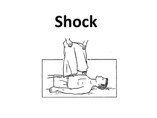
SHOCK

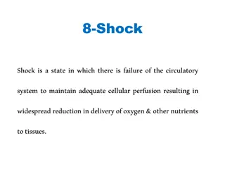
Shock is a life-threatening condition characterized by inadequate delivery of oxygen and nutrients to vital organs relative to their metabolic demand. Samuel D. Gross defined shock as the rude unhinging of the machinery of life. It is a transition between life and death, with mortality rates exceeding 20%. Shock can be classified into categories such as Hematologic, Neurologic, Vasogenic, and Cardiogenic. Causes of shock may require primarily the infusion of volume, such as Hemorrhagic shock, Traumatic shock, Gastrointestinal shock, and Hypovolemia. Learn about the vital signs, symptoms, and pathophysiology of shock's stages - Compensated and Decompensated.

Download Presentation

Please find below an Image/Link to download the presentation.
The content on the website is provided AS IS for your information and personal use only. It may not be sold, licensed, or shared on other websites without obtaining consent from the author.If you encounter any issues during the download, it is possible that the publisher has removed the file from their server.
You are allowed to download the files provided on this website for personal or commercial use, subject to the condition that they are used lawfully. All files are the property of their respective owners.
The content on the website is provided AS IS for your information and personal use only. It may not be sold, licensed, or shared on other websites without obtaining consent from the author.
E N D
Presentation Transcript
What is shock ? Samuel D. Gross defined shock as the rude unhinging of the machinery of life.
What is shock ? A transition between life and death
What is shock ? Can we define shock as hypotension?
What is shock ? Shock is defined as a life-threatening condition characterized by inadequate delivery of oxygen and nutrients to vital organs relative to their metabolic demand mortality rates exceed 20%
Classification Categories of shock ?
Classification Hematologic Neurologic Vasogenic Cardiogenic
Classification Causes That Require Primarily the Infusion of Volume
Classification Hemorrhagic shock Traumatic Gastrointestinal Body cavity Hypovolemia Gastrointestinal losses Dehydration from insensible losses Third-space sequestration from inflammation
Stage Vital Signs Signs and Symptoms Pathophysiology Normal blood pressure Normal to slightly heart rate Tachypnea Delayed capillary refill Cool hands and feet Pale mucous membranes Restlessness, anxiety Oliguria Vasoconstriction maintains blood flow to essential organs, but tissue ischemia occurs in less essential areas Compensated Blood pressure decreasing Tachycardic >120/min Tachypneic > 30-40/min Waxen, cool, clammy skin Pale or cyanotic mucus membranes Profound weakness Metabolic (lactic) acidosis Anxiety Absent or peripheral pulses Blood pressure decreases as the vascular tone decreases. Dysfunction to all organs is imminent. Anaerobic metabolism ensues, causing lactic acidosis. Decompensated Profound hypotension Lactate > 8mEq/L Metabolic acidosis causes postcapillary sphincters to open and release stagnant and coagulated blood. Excessive potassium and acid causes dysrhythmias. Cellular damage is irreversible. Irreversible
Classification Causes That Require Improvement in Pump Function by Either Infusion of Inotropic Support or Reversal of the Cause of Pump Dysfunction
Classification Myocardial ischemia Coronary artery thrombosis Arterial hypotension with hypoxemia Cardiomyopathy Acute myocarditits Chronic diseases of heart muscle (ischemic, diabetic, infiltrative, endocrinologic, congenital) Cardiac rhythm disturbances Atrial fibrillation with rapid ventricular response Ventricular tachycardia Supraventricular tachycardia
Classification Hypodynamic septic shock Overdose of negative inotropic drug Beta-blocker Calcium channel antagonist overdose Structural cardiac damage Traumatic (e.g., flail mitral valve) Ventriculoseptal rupture Papillary muscle rupture
Classification Causes That Require Volume Support and Vasopressor Support
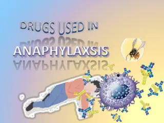
Classification Hyperdynamic septic shock Anaphylactic shock Central neurogenic shock Drug overdose
Classification Problems That Require Immediate Relief from Obstruction to Cardiac Output
Classification Pulmonary embolism Cardiac tamponade Pneumothorax Valvular dysfunction Acute thrombosis of prosthetic valve Critical aortic stenosis Congenital heart defects in newborn (e.g., closure of patent ductus arteriosus with critical aortic coarctation) Critical idiopathic subaortic stenosis (hypertrophic obstructive cardiomyopathy)
Search for: 1. Hemorrhagic shock 2. Tension pneumothorax 3. Cardiac tamponade 4. Cardiac injury History of trauma? YES O N Evidence of gastrointestinal hemorrhage, vomiting, or diarrhea? YES Volume resuscitate NO 1. Begin treatment for sepsis syndrome 2. Search for source of infection 3. Consider drawing thyroid function tests Fever or hypothermia? YES NO 1. Treat for cardiogenic shock from myocardial ischemia 2. Consider massive pulmonary embolism with right ventricular strain effect Electrocardiographic evidence for ischemia or chest pain with major risk factors for coronary artery disease? YES
1. Evaluate or treat for ingestion of negative inotropic drug Draw thyroid function tests Consider treatment for addisonian crisis or steroid withdrawal Unexplained bradycardia with hypotension? yes 2. 3. NO Unexplainedhypoxemia? Rule out pulmonary embolism YES NO 1. 2. Volume resuscitate Emergent abdominal computed tomography or surgical consultation to evaluate for peritoneal inflammation or vascular rupture YES Abdominal or low back pain? NO Wheezing with hives or skin flushing? YES Treat for anaphylaxis
EARLY DETECTION OF SHOCK Why is it important?
Quantitative Resuscitation Resuscitating patients to predefined physiologic endpoints indicating that systemic perfusion and vital organ function have been restored first described in 1988
Quantitative Resuscitation significantly reduce mortality and morbidity rates in ED patients with severe sepsis or septic shock (early goal directed therapy) importance of the first 6 hours of resuscitation.
Monitoring Perfusion Status The basics: Electrocardiography pulse oximetry BP
Monitoring Perfusion Status Arterial pressure monitoring line Accurate When using vasoactive agents Central venous pressure 8-12 (10-15 when ventilated) Goal-directed resuscitation Improves outcome in patients with septic shock Vasoactive agents Urine output 1 mL/kg/hr in persons without renal disease
Monitoring Perfusion Status pulmonary artery catheter remains controversial Insufficient data have been published to support the use or avoidance of PACs in ED populations 5 RCT s investigating the management of critically ill patients with a PAC ,none have found a benefit in terms of survival or length of stay
Ventilation RSI is the preferred method of airway control in most patients with shock
Hemorrhagic Shock Ensure adequate ventilation/oxygenation Provide immediate control of hemorrhage, when possible (e.g., traction for long bone fractures, direct pressure) Initiate judicious infusion of isotonic crystalloid solution (10 20 mL/kg) With evidence of poor organ perfusion and 30- minute anticipated delay to hemorrhage control, begin packed red blood cell (PRBC) infusion (5 10 mL/kg)
Hemorrhagic Shock With suspected central nervous system trauma or Glasgow Coma Scale score <9, immediate PRBC transfusion may be preferable as initial resuscitation fluid Treat coincident dysrhythmias (e.g., atrial fibrillation with synchronized cardioversion)
Cardiogenic Shock Ameliorate increased work of breathing; provide oxygen and positive end-expiratory pressure (PEEP) for pulmonary edema Begin vasopressor or inotropic support; norepinephine (0.5 mg/min) and dobutamine (5 mg/kg/min) are common empirical agents
Cardiogenic Shock Seek to reverse the insult (e.g., initiate thrombolysis, arrange percutaneous transluminal angioplasty) Consider intra-aortic balloon pump counterpulsation for refractory shock
Systemic Inflammatory Response Syndrome (SIRS) Two or more of the following: 1. Temperature >38 C or <36 C 2. Heart rate >90 beats/min 3. Respiratory rate >20 breaths/min or Paco2 <32 mm Hg 4. While blood cell count >12,000/mm3, <4000/mm3, or >10% band neutrophilia
Severe Sepsis SIRS with suspected or confirmed infection and associated with organ dysfunction or hypotension; organ dysfunction may include presence of lactic acidosis, oliguria, or altered mental status
Septic Shock SIRS with suspected or confirmed infection with hypotension despite adequate fluid resuscitation; septic shock should still be diagnosed if vasopressor therapy has normalized blood pressure
Summary Ensure adequate oxygenation; remove work of breathing Administer 20 mL/kg of crystalloid
Summary Begin antimicrobial therapy; attempt surgical drainage or de bridement Begin PRBC infusion for hemoglobin < 8 g/dL If volume restoration fails to improve organ perfusion, begin vasopressor support; initial choice includes dopamine, infused at 5 15 mg/kg/min, or norepinephrine, infused at 0.5 mg/min