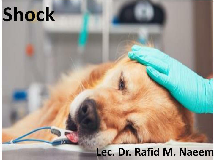
Shock: Types, Causes, and Responses
Shock is a clinical condition where tissue oxygen delivery is compromised, leading to tissue hypoxia. This article explores the classification of shock into hypovolemic, cardiogenic, distributive, and hypoxic types, along with their causes and manifestations. Learn about the body's compensatory mechanisms to preserve vital organ function in response to shock.
Download Presentation

Please find below an Image/Link to download the presentation.
The content on the website is provided AS IS for your information and personal use only. It may not be sold, licensed, or shared on other websites without obtaining consent from the author. If you encounter any issues during the download, it is possible that the publisher has removed the file from their server.
You are allowed to download the files provided on this website for personal or commercial use, subject to the condition that they are used lawfully. All files are the property of their respective owners.
The content on the website is provided AS IS for your information and personal use only. It may not be sold, licensed, or shared on other websites without obtaining consent from the author.
E N D
Presentation Transcript
Shock Lec. Dr. Rafid M. Naeem
Shock is the clinical picture observed when tissue oxygen delivery or utilization is compromised. Oxygen delivery (DO2) depends upon adequate cardiac output (CO) and arterial oxygen content (CaO2) Tissue hypoxia is the result of inadequate oxygen delivery or utilization .
The body responds to tissue hypoxia or shock by compensatory mechanisms to preserve vital organ function and host viability. . These compensatory mechanisms are manifest as the classic clinical findings in a patient in shock: 1. tachycardia (to increase oxygen delivery), 2. tachypnea (to increase oxygenation), 3. peripheral vasoconstriction (to maintain perfusion of vital organs), and 4. mental depression (in response to decreased perfusion or hypoxia).
Shock can be classified according to its main contribution to impaired tissue oxygenation Hypovolemic shock Cardiogenic shock Distributive shock Hypoxic shock
1. Hypovolemic shock occurs when circulating volume is inadequate. . Hypovolemic shock is a consequence of a reduction in the circulating intravascular volume. It leads to impaired oxygen delivery through a reduction in venous return to the heart (preload) and, as a consequence, reduced cardiac output. ( . . ) Hypovolemic shock can be caused by hemorrhagic losses (internal or external bleeding) or by the loss of other body fluids (third space, gastrointestinal/urinary losses, burns). ( ( ) / .
2. Cardiogenic shock is used to describe conditions that impair forward flow of blood from the heart. Although sometimes described as obstructive shock, conditions that restrict right ventricular filling may also be grouped under cardiogenic shock. " " . Cardiogenic shock results from an inability of the heart to propel the blood through the circulation. Cardiogenic shock can result from anything that interferes with the ability of the heart to fill (diastolic failure) or pump blood (systolic failure). . ( .) . ) (
3. Distributive shock is caused by inappropriate vasodilation, resulting in inadequate effective circulating volume. Distributive shock is characterized by an impairment of the mechanisms regulating vascular tone, with maldistribution of the vascular volume and massive systemic vasodilation. . The consequence of this decrease in systemic vascular resistance is that the amount of blood in the circulation is inadequate to fill the vascular space, creating relative hypovolemia and a reduction in venous return. .
The most common causes of distributive shock are sepsis and the systemic inflammatory response syndrome (SIRS). (SIRS) However, distributive shock can be caused by anaphylactic reactions (anaphylactic shock), drugs (anesthetics), or severe damage to the central nervous system associated with sudden loss of autonomic nervous stimulation on the vessels (neurogenic shock). ( ) ) ) ( (
4. Hypoxic shock results from inadequate arterial oxygen content or impaired mitochondrial function (impaired oxygen uptake) ( ) Hypoxic shock is characterized by adequate tissue perfusion but inadequate arterial oxygen content or cellular oxygen utilization.
The most common causes of hypoxic shock are anemia (reduced hemoglobin [Hb] concentration anemic hypoxia) and hypoxemia (reduced PaO2 or SaO2 hypoxemic hypoxia), associated with respiratory failure. Arterial hemoglobin oxygen saturation (SaO2) is a measurement of the percentage of how much hemoglobin is saturated with oxygen. ( . SaO 2 ( Arterial oxygen partial pressure (PaO2). This measures the pressure of oxygen dissolved in the blood and how well oxygen is able to move from the airspace of the lungs into the blood. 2 ( (. PaO .
Hypoxic shock can also be associated with toxicities that impair the ability of hemoglobin to bind oxygen, such as methemoglobinemia (e.g., acetaminophen toxicity in cats) or carbon monoxide poisoning. ( ) . In metabolic or cytopathic shock, despite adequate tissue levels of oxygen, cells are not able to produce a sufficient amount of energy. This form of hypoxic shock is caused by intracellular interference with oxygen uptake and aerobic energy production (e.g., sepsis, toxins). . ) (
Treatment Initial stabilization should focus on airway, breathing, circulation, and neurologic derangements. Major diagnostics often need to be delayed until the patient is stabilized. ) . . (
Major components of therapy for patients in shock rely on supportive care : 1. Optimization of oxygen delivery 2. reestablishment of adequate tissue perfusion and aggressive support of organ function 3. Identification and treatment of the underlying/initiating disease .1 .2 .3
In veterinary practice; indirect indications related to end-organ perfusion and intravascular volume status are considered more practical endpoints: . : These markers include traditional vital signs and hemodynamic variables (heart rate, capillary refill time, urinary output, blood pressure) . (
Oxygen supplementation: Flow-by oxygen, the simplest method, maintains a high-flow source of humidified oxygen close to the patient s mouth or nose. Fluid therapy in shock patients is based on the rapid administration of large volumes of intravenous fluid to replenish the relative or absolute intravascular volume deficit and restore perfusion Hypothermia is frequently present in shock states. Preventing heat loss and administering warmed fluids are the safest approaches to temperature correction.
it is preferred feeding the patient because it supports gastrointestinal integrity, thus minimizing bacterial translocation. Most patients will benefit from some level of analgesia and/or sedation to reduce the stress associated with pain and hospitalization (e.g., trauma, cardiogenic shock). /
Therapies designed to address the underlying cause of shock should be established as soon as possible. These include, for example hemostasis of bleeding in hypovolemic shock, and infection control in patients with septic shock. .
Be serious and study hard
