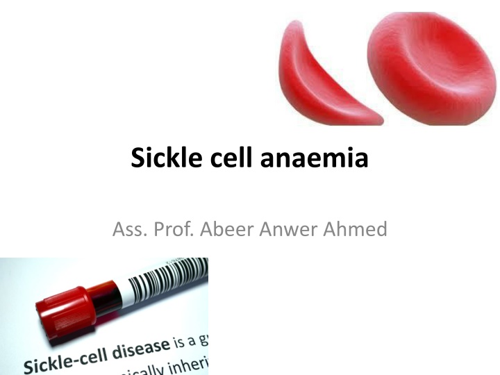
Sickle cell anaemia
Sickle cell disease is a group of hemoglobin disorders caused by a genetic abnormality leading to misshapen red blood cells, affecting predominantly regions like sub-Saharan Africa, the Mediterranean Basin, the Middle East, and India. The disease is prevalent in these areas due to the protective effect against severe malaria provided by the sickle cell trait. This inherited condition results from the substitution of valine for glutamic acid in the hemoglobin chain, leading to the polymerization of deoxygenated sickle hemoglobin into long fibers that can block blood flow, causing organ damage. Globally, a significant number of babies are born with hemoglobinopathies each year, with a high percentage in Nigeria. The Middle East, including countries like Saudi Arabia and Iraq, also faces a significant prevalence of sickle cell disease due to a high rate of consanguineous marriages affecting the distribution of the disease clusters.

Download Presentation

Please find below an Image/Link to download the presentation.
The content on the website is provided AS IS for your information and personal use only. It may not be sold, licensed, or shared on other websites without obtaining consent from the author.If you encounter any issues during the download, it is possible that the publisher has removed the file from their server.
You are allowed to download the files provided on this website for personal or commercial use, subject to the condition that they are used lawfully. All files are the property of their respective owners.
The content on the website is provided AS IS for your information and personal use only. It may not be sold, licensed, or shared on other websites without obtaining consent from the author.
E N D
Presentation Transcript
Sickle cell anaemia Ass. Prof. Abeer Anwer Ahmed
Sickle cell disease is a group of haemoglobin disorders resulting from the inheritance of the sickle globin gene. Sickle cell disease (SCD) is an autosomal recessive
The sickle globin abnormality is caused by substitution of valine for glutamic acid in position 6 in the chain
Hb S(Hb 22S) is insoluble and forms crystals when exposed to low oxygen tension Deoxygenated sickle haemoglobin polymerizes into long fibres, each consisting of seven intertwined double strands with cross linking The red cells sickle and may block different areas of the microcirculation or large vessels causing infarcts of various organs.
Epidemiology The prevalence of the SCD is high in sub-Saharan Africa, Mediterranean Basin, Middle East and India because of the level of protection that the sickle cell trait provides against severe Malaria.
According to the WHO, annually over 330,000 babies are born with hemoglobinopathies (83 % with SCD and 17% with Thalassemia). Around 5.2% of the world s population carry the genes of hemoglobinopathies, 2.28% have mutations for SCA, with the vast majority presents in Nigeria.
India has been reported as the second worst affected country of predicted SCA births, with frequency of sickle cell trait reaches level as high as ~ 40%. Hemoglobinopathies are common hematologic disorders in the Arab world due to high level of consanguineous marriages. In Saudi Arabia, the consanguineous marriages are about (40-50%). (17)The prevalence rate for SCD was 49.6% (45.8% for the trait and 3.8% for the disease).
In Iraq, the distribution of the disease clusters in two main regions one in the south (Basra) where the frequency of sickle cell trait is about 6.48%,(19,20)and the other is in the north among Kurdish population where the prevalence of sickle cell carrier state is about 1.2%.
genotypes of Sickle cell disease denotes all SCD containing at least one sickle gene, in which HbS makes at least half of the total hemoglobin present. HbSS disease (Sickle Cell Anemia): is the most common type and usually present with moderate or severe symptoms -
- HbS/0thalassemia (compound heterozygosity for HbS and 0 thalassemia): clinically, it is indistinguishable from sickle cell anemia. - HbS/ +thalassemia: is characterized by mild to moderate disease.
HbSC disease (compound heterozygosity for HbS and HbC): is characterized by moderate disease severity. - HbS/hereditary persistence of fetal Hb (S/HPFH): is characterized by very mild or even asymptomatic phenotype. - HbS/HbE syndrome: very rare with a phenotype similar to HbS/ +thalassemia.
- HbSS/ -Thalassemia: concurrent -thalassemia with SCA, present with SCA symptoms but milder, symptoms are ameliorated due to less formation of HbS, as a result of low -globin availability and thus sickling is reduced. - There are rare combinations of HbS with other abnormal hemoglobin, such as HbD Los Angeles, HbO Arab, G-Philadelphia, and others.
Combination of haemoglobin S with other geneticdefects of haemoglobin The most common of these are Hb S/ thalassaemia, and sickle cell/C disease. In Hb S/ thalassaemia, the MCV and MCH are lower than in homozygous Hb SS. The clinical picture is of sickle cell anaemia; splenomegaly is usual. Patients with HbSC disease have a particular tendency to thrombosis and pulmonary embolism, especially in pregnancy. In general, when compared with Hb SS disease, they have a higher incidence of retinal abnormalities, milder anaemia, splenomegaly and generally a longer life expectancy. Diagnosis is made by haemoglobin electrophoresis or HPLC, particularly with family studies.
Clinical features Homozygous disease severe haemolytic anaemia punctuated by crises. The symptoms of anaemia are often mild in relation to the severity of the anaemia because Hb S gives up oxygen (O2) to tissues relatively easily compared with Hb The clinical expression of Hb SS is very variable, some patients an almost normal life, free of crises, but others develop severe crises even as infants and may die in early childhood or as young adults
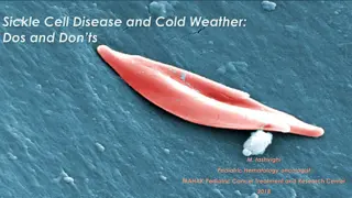
Vasoocclusive crises Painful These are the most frequent. precipitated by :infection, acidosis, dehydration or deoxygenation (e.g. altitude, operations, obstetric delivery, stasis of the circulation, exposure to cold, violent exercise). Infarcts causing severe pain occur in the bones (hips, shoulders and vertebrae are commonly affected) The hand foot syndrome (painful dactylitis caused by infarcts of the small bones) is frequently the first presentation of the disease and may lead to digits of varying lengths
avascular necrosis with flattening of the femoral heads, more marked on the right,coarsening of the bone architecture and cystic areas in the right femoral neck caused by previous infarcts.
Visceral These are caused by sickling within organs causing infarction and pooling of blood, often with a severe exacerbation of anaemia. The acute sickle chest syndrome is the most common cause of death both in children and adults. It presents with dyspnoea, falling arterial PO2, chest pain and pulmonary infiltrates on chest X ray.
Splenic sequestration is typically seen in infants and presents with an enlarging spleen, falling haemoglobin and abdominal pain. Treatment is with transfusion. Attacks tend to be recurrent and splenectomy is often needed. Priapism and liver and kidney damage due to repeated small infarcts are other complications
Aplastic crises These occur as a result of infection with parvovirus or from folate deficiency and are characterized by a sudden fall in haemoglobin and reticulocytes, usually requiring transfusion
Haemolytic crises These are characterized by an increased rate of haemolysis and fall in haemoglobin but rise in reticulocytes and usually accompany a painful crisis
organ damage The most serious is of the brain (a stroke occurs in 7% of all patients) or spinal cord. Up to a third of children have had a silent cerebral infarct by the age of 6 years. Ulcers of the lower legs are common, as a result of vascular stasis and local ischaemia
The spleen is enlarged in infancy and early childhood but later is often reduced in size as a result of infarcts (autosplenectomy). Pulmonary hypertension Infections are frequent partly due to hyposplenism. Pneumonia, urinary tract infections and Gram negative septicaemia are common. Osteomyelitis may also occur, usually from Salmonella spp.
Laboratory findings 1 The haemoglobin is usually 60 90 g/L low in comparison to mild or no symptoms of anaemia. 2 Sickle cells and target cells occur in the blood . Features of splenic atrophy (e.g. Howell Jolly bodies) may also be present. 3 Screening tests for sickling are positive when the blood is deoxygenated (e.g. with dithionate and Na2 HPO4). 4 HPLC or haemoglobin electrophoresis :in Hb SS, no Hb A is detected. The amount of Hb F is variable and is usually 5 15%, larger amounts are usually associated with a milder disorder.
Sickle cell anaemia: peripheral blood film showing deeply staining sickle cells, target cells and polychromasia.
Treatment 1 Prophylactic avoid those factors known to precipitate crises, especially dehydration, anoxia, infections, stasis of the circulation and cooling of the skin surface. 2 Folic acid (e.g. 5 mg once weekly). 3 Good general nutrition and hygiene. 4 Pneumococcal, Haemophilus and meningococcal vaccination and regular oral penicillin are effective at reducing the infection rate with these organisms. Oral penicillin should start at diagnosis and continue at least until puberty. Hepatitis B vaccination is also given as transfusions may be needed. 5 Crises treat by rest, warmth, rehydration by oral fluids and/or intravenous normal saline (3 L in 24 h) and antibiotics if infection is present. Analgesia at the appropriate level should be given.
Blood transfusion is given only if: there is very severe anaemia with symptoms. Exchange transfusion may be needed, particularly if there is neurological damage, a visceral sequestration crisis or repeated painful crises. This is aimed at achieving an Hb S percentage of less than 30% in severe cases and, after a stroke, is continued for at least 2 years.
6 Particular care is needed in pregnancy and anaesthesia. There is debate as to whether or not patients need transfusions with normal blood to reduce Hb S levels during pregnancy or before delivery or for minor operations. Routine transfusions throughout pregnancy are given to those with a poor obstetric history or a history of frequent crises. Careful anaesthetic and recovery techniques must be used to avoid hypoxaemia or acidosis
7 Transfusions these are also sometimes given repeatedly as prophylaxis to patients having frequent crises or who have had major organ damage (e.g. of the brain) or show abnormal transcranial Doppler studies. The aim is to suppress Hb S production over a period of several months or evenyears. Iron overload, best assessed by the total number of units transfused and liver iron (and may need iron chelation therapy), and alloimmunization against donated blood are common problems
8 Hydroxycarbamide can increase Hb F levels and improves the clinical course of children or adults. It is given to those with severe or moderately severe disease, e.g. who are having three or more painful crises each year. It should not be used during pregnancy. 9 Stem cell transplantation can cure the disease, with 80% disease free. The mortality rate is less than 10%. Transplantation is only indicated in the severest of cases whose quality of life or life expectancy are substantially impaired. 10 Research into other drugs (e.g. butyrates) to enhance Hb F synthesis or to increase the solubility of Hb S is taking place
Sickle cell trait This is a benign condition with no anaemia and normal appearance of red cells in a blood film. Haematuria is the most common symptom and is thought to be caused by minor infarcts of the renal papillae. Hb S varies from 25 45% of the total haemoglobin Care must be taken with anaesthesia, pregnancy and at high altitudes.
Haemoglobin C disease This genetic defect of haemoglobin is frequent in West Africa and is caused by substitution of lysine for glutamic acid in globin chain at the same point as the substitution in Hb S. Hb C tends to form rhomboidal crystals and in the homozygous state there is a mild haemolytic anaemia with marked target cell formation, cells with rhomboidal shape and microspherocytes The spleen is enlarged. The carriers show a few target cells only.
Homozygous Hb C disease: peripheral blood film showing many target cells, deeply staining rhomboidal and
Haemoglobin D disease This is a group of variants all with the same electrophoretic mobility. Heterozygotes show no haematological abnormality while homozygotes have a mild haemolytic anaemia. Haemoglobin E disease This is the most common haemoglobin variant in South East Asia. In the homozygous state, there is a mild microcytic, hypochromic anaemia. Haemoglobin E/ 0 thalassaemia, however, resembles homozygous 0 thalassaemia both clinically and haematologically


 is insoluble and")
 is insoluble and")


 in a")