Special Staining Techniques for Bacterial Identification
Learn about special staining techniques like Zeihl-Neelson (acid-fast stain) and Spore stain used to visualize bacteria structures such as spores, capsules, and flagella. Understand the procedures and importance of these staining methods in identifying bacteria for diagnostic purposes.

Download Presentation

Please find below an Image/Link to download the presentation.
The content on the website is provided AS IS for your information and personal use only. It may not be sold, licensed, or shared on other websites without obtaining consent from the author. If you encounter any issues during the download, it is possible that the publisher has removed the file from their server.
You are allowed to download the files provided on this website for personal or commercial use, subject to the condition that they are used lawfully. All files are the property of their respective owners.
The content on the website is provided AS IS for your information and personal use only. It may not be sold, licensed, or shared on other websites without obtaining consent from the author.
E N D
Presentation Transcript
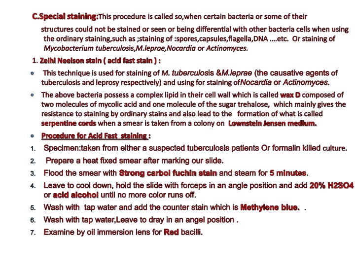
C.Special staining:This procedure is called so,when certain bacteria or some of their structures could not be stained or seen or being differential with other bacteria cells when using the ordinary staining,such as ;staining of :spores,capsules,flagella,DNA . etc. Or staining of Mycobacterium tuberculosis,M.leprae,Nocardia or Actinomyces. 1. Zeihl Neelson stain ( acid fast stain ) : This technique is used for staining of M. tuberculosis &M.leprae (the causative agents of tuberculosis and leprosy respectively) and using for staining ofNocardia or Actinomyces. The above bacteria possess a complex lipid in their cell wall which is called wax D composed of two molecules of mycolic acid and one molecule of the sugar trehalose, which mainly gives the resistance to staining by ordinary stains and also lead to the formation of what is called serpentine cords when a smear is taken from a colony on Lownstein Jensen medium. Procedure for Acid Fast staining : Specimen:taken from either a suspected tuberculosis patients Or formalin killed culture. Prepare a heat fixed smear after marking our slide. Flood the smear with Strong carbol fuchin stain and steam for 5 minutes. Leave to cool down, hold the slide with forceps in an angle position and add 20% H2SO4 or acid alcohol until no more color runs off. Wash with tap water and add the counter stain which is Methylene blue. . Wash with tap water,Leave to dray in an angel position . Examine by oil immersion lens for Red bacilli.
2. Spore stain : During sporulation process,the cell forms several layers : Core: which contains its DNA and some ribosomes and dipicolinic acid and some other most important factors that keep the cell resistant to most adverse conditions. Cortex: consist of double layer of peptidoglycan, differs from that of the cell wall. Exosporium: consist of keratinized protein. The above layers give the spore protection against several hard environmental conditions, at the same time, become difficult to stain the spore with the ordinary staining techniques; therefore its staining requires a special procedure such as steaming in order to allow the stain to penetrate into the spore. Procedure for Spore staining ( Schaeffer-fulton mfthod) : Prepare a heat fixed smear of a spore forming bacteria such as Bacillus subtilis or any species of Clostridium after marking our slide. Flood the smear with Malachite green stain and steam for 5 minutes. Leave to cool down and wash with tap water. Add Safranine as a counter stain ,for 1 minutes. Wash with tap water, Leave to dray in upright position . Examine by oil immersion lens . Location and the size of endospores vary with species,thus they are of value in identifying bacteria. Endospores position within the cell is characteristic. It can be either : 1.Central. 2. Subterminal. 3. Terminal.
Endospores: Endospores are bright green. Vegetative Cells: Vegetative cells are brownish red to pink. Spores may be located in the middle of the cell, at the end of the cell, or between the en
3. Capsule stain : Capsule is a gelatinous spherical or oval shape surrounding certain of bacteria as a protective extracellular structure, protect the cell from phagocytosis by phagocytic cell, Therefore this structure is considered, in several species of bacteria, as a virulence factor, and its loss leads to the loss of the virulence in that bacteria, also it is Antigenic . The capsule is consisting of 97-98% water and only 2-3% of dissolved solid.These solid differ with the different species,may be consisting of polysaccharides,glycoproteins , polysaccharides with certain lipids .. etc. Since the capsule consisting of large amount of water, it requires a special procedure for staining . One of the procedure is called Hiss method : Procedure for Capsule staining (Hiss method ) : Prepare a smear for bacteria that possess a capsule,such as; Klebsiella or Pneumococci and heat fix very gently on the flame to avoid damaging of the capsule by excess heat. Flood the smear with 1% aqueous solution of crystal violet stain. Steam very gently for 2-3 minutes. Leave to cool and rins with 20% copper sulfate ( CuSo4 ). Leave to dray and examine under the oil immersion lens.capsule appear faint surrounding dark blue cell. Other methods for view capsule : 1- Capsule-swelling test ( Quelling reaction ). 2- Negative stain : India ink , Nigrosin.
Another procedure: staining with India ink: 1)Place a loop-full of capsulated bacteria on one end of the slide. 2)Add a drop of India ink on the bacteria. 3)With another slide withdraw the mixture to make a thin film. 4)Allow to dry, and examine under the oil immersion.
The capsule appears as a clear zone between the refractile cell outline and the dark background. Positive control Klebsiella pneumoniae (ATCC e
4. Flagella : Tiny hair like organism of locomotion presence of flagella ; numbers and patterns of arrangement provide clues to identify species. Flagellar stain : Flagellar stain contains Rosaniline dyes and a mordant,applied to bacterial suspension fixed in formalin and spread across a glass slide . The formalin fixes flagella, the dye and mordant then precipitate around these fixed surface, enlarging their diameter and making then visible. The mordant is tannic acid and the dye is basic fuchsin. Flagella can be viewed by: E. M. D.F.E ( Dark field ). Flagellar staining.

 IN TISSUE")
 IN TISSUE")