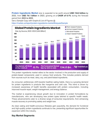
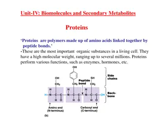
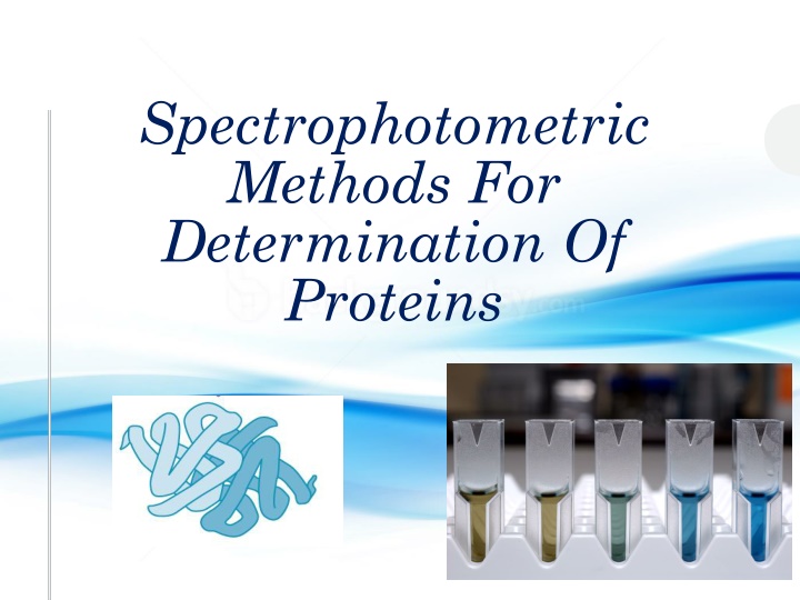
Explore spectrophotometric methods such as the Bradford Method and Warburg-Christian Method for determining protein concentration in the lab. Learn about the importance of protein assays in life science research and the factors to consider when choosing a method.
Please find below an Image/Link to download the presentation.
The content on the website is provided AS IS for your information and personal use only. It may not be sold, licensed, or shared on other websites without obtaining consent from the author. If you encounter any issues during the download, it is possible that the publisher has removed the file from their server.
You are allowed to download the files provided on this website for personal or commercial use, subject to the condition that they are used lawfully. All files are the property of their respective owners.
The content on the website is provided AS IS for your information and personal use only. It may not be sold, licensed, or shared on other websites without obtaining consent from the author.
Objectives To Learn Different Method Of proteins determination In this Lab you will using the following spectrophotometric methods: 1. Bradford Method. Chemical reagents are added to the protein solutions to develop a color whose intensity is measured in a spectrophotometer. 2. Warburg-Christian Method ( A280/ A260 Method). Relies on direct spectrophotometric measurement
Important Terms Assays Qualitative assays Quantitative assays Determine the concentration of a substance Determine if specific substance is there or not, by color or some other quality Specifity and sensivity Specificity of an assay relates to how good the assay is in discriminating between the requested analyte and interfering substances Sensitivity of an assay is a measure of how little of the analyte the method can detect
Importance of determining concentration of protein Protein assays are one of the most widely used methods in life science research. Estimation of protein concentration is necessary cell biology, molecular biology and other research applications. Is necessary before processing protein samples for isolation, protein purification, separation and analysis.
The determination of protein concentration Spectrophotometric and colorimetric methods Acid hydrolyse a portion of the sample Used for routine estimation, most of them are colometric And then carry out amino acid analysis on the hydrolysate A truly accurate method Where a portion of the protein solution is reacted with a reagent that produces a coloured product. However, this is expensive and relatively consuming, particularly multiple samples are to be analysed. time- if However, none of these methods is absolute,
Spectrophotometric method for determining the protein concentration Warburg christian Bicinchoninic acid method lowry Bradford (A280/A260) There are a wide variety of protein assays available. but each assay has its own advantages and limitations The factors that you should consider to choose Method : Sensitivity The presence of interfering substance Time available of the assay Cost
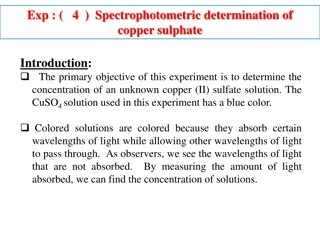
1-Bicinchoninic acid method In general, this method has a high sensitivity (1 g ) Principle: Cu+2 form a complex with nitrogen of the peptide bond under alkaline conditions producing cu+(the Cu++ was reduced to Cu+) This cu+ will then chelated by BCA to produce a copper-BCA complex with maximum absorbance 562 nm Alkaline condition
2-Bradford assay Fast, accurate and high sensitivity [1 g protein can be detected]. The method is recommended for general use, especially for determining protein content of cell fractions and assessing protein concentrations for gel electrophoresis, about 1- 20 g protein for micro assay or 20-200 g protein for macro assay. Principle: Generally, Coomessie brilliant blue G-250 bind to protein (binds particularly to basic and aromatic amino acids residues ) in acidic solution Make a complex which will shift the wavelength of maximum absorbance ( max) 465 to 595 nm. Cell fractionation
2-Bradford assay How it happened? Coomessie brilliant blue G-250 dye exists in three forms: cationic (red), neutral (green), and anionic (blue) Under acidic conditions, the dye is predominantly in the doubly protonated red cationic form (Amax = 470 nm). However, when the dye binds to protein, it is converted to a stable unprotonated blue form (Amax = 595 nm) It is this blue protein-dye form that is detected at 595 nm This complex stabilized by hydrophobic and ionic interaction
Bradford assay Bradford reagent alone maximum absorbance at (465 nm) Bradford reagent and protein maximum absorbance at (595 nm)
Bradford assay-Method A- Set up 9 tubes and label them as follows: Bovine Serum Albumin(BSA) (150 g/ml) Tube Distilled Water Unknown Concentration ( g/ml) - 1 ml - Blank (blank) 0.07 ml 0.93 ml - 10.5 A 0.13 ml 0.87 ml - 19.5 B 0.26 ml 0.74 ml - 39 C 0.4 ml 0.6 ml - 60 D 0.66 ml 0.34 ml - 99 E 1 ml - - 150 F - - 1 ml ? G - - 1 ml ? H Add 5ml of Bradford reagent to each tube [blank H]. C- Mix and Incubate at room temperature for 5 min. D- Measure the absorbance at 595 nm.
Bradford assay-Results Tube Concentration ( g/ml) Absorbance at 595 nm Blank (blank) 10.5 A 19.5 B 39 C 60 D 99 E 150 F =.................. G =.................. H
3-Warburg christian (A280/A260) Relies on direct spectrophotometric measurement. Fast Semiquantitative analysis Principle: Proteins can absorb light at 280 ultraviolet This is because proteins contains aromatic amino acids tyrosine and tryptophan give proteins . The amount of these residues vary greatly from protein to protein so this method is semiquantitave
Warburg christian (A280/A260) Nucleic acid interfere with this method. So to solve this problem, we will measure the absorbance at 280 then we measure at 260 Calculate A280/A260 ratio, then from a specific table we can get the correction factor Concentration=A280 x correction factor Or by another way: [groves formula]: Protein concentration [mg/ml]=[1.55 X A280]-[0.76 X A260]
Warburg christian (A280/A260) -Calculate the protein concentration in the unknown from the following equations: A280 = A260 = A280/ A260 = Correction factor = A280 x correction factor = mg/ml protein Unknown concentration = mg/ml 2-or [groves formula]: Protein concentration [mg/ml]=[1.55 X A280]-[0.76 X A260]
Warburg christian (A280/A260) -A protein solution that has a high A280/A260 ratio: Less contaminated by DNA. [It shows a lower absorbance at 260nm comparing to absorbance at 280nm]. -A protein solution that has a low A280/A260 ratio: Highly contaminated by DNA. [It shows a higher absorbance at 260nm comparing to absorbance at 280nm].
Summary Protein assay is important in many aspects There are Many Methods for protein determination, each had it own advantages and disadvantages ASSAY ABSORPTION MECHANISM reagent Tyrosine and tryptophan absorption UV absorption 280 nm No reagent copper reduction (Cu2+ to Cu1+), BCA reaction with Cu1+ Bicinchoninic acid 562 nm BCA complex formation between Coomassie brilliant blue dye and proteins Bradford or Coomassie brilliant blue Coomassie brilliant blue 595 nm
References Principles and Techniques of Biochemistry and Molecular Biology Quick Start Bradford Protein Assay Instruction Manual


")