Spicules in Sponge Taxonomy
Spicules, microscopic crystalline structures found in sponges, play a crucial role in providing structural support and defense against predators. Learn about the types, classification, and functions of spicules in sponge taxonomy.

Download Presentation

Please find below an Image/Link to download the presentation.
The content on the website is provided AS IS for your information and personal use only. It may not be sold, licensed, or shared on other websites without obtaining consent from the author.If you encounter any issues during the download, it is possible that the publisher has removed the file from their server.
You are allowed to download the files provided on this website for personal or commercial use, subject to the condition that they are used lawfully. All files are the property of their respective owners.
The content on the website is provided AS IS for your information and personal use only. It may not be sold, licensed, or shared on other websites without obtaining consent from the author.
E N D
Presentation Transcript
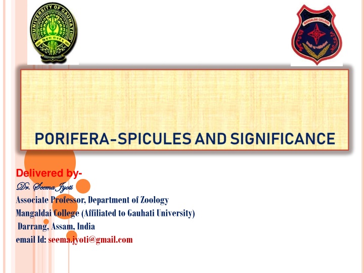
Delivered by- Dr. Seema Jyoti Dr. Seema Jyoti Associate Professor, Department of Zoology Mangaldai College (Affiliated to Gauhati University) Darrang, Assam, India email Id: seema.jyoti@gmail.com
INTRODUCTION Spicules elements found in most sponges. They provide structural support and deter predators. Large spicules that are visible to the naked eye are referred to as smaller, microscopic ones are termed microscleres. The meshing of many sponge s skeleton and thus it provides structural support and defence composition, size, and shape of spicules is one of the largest determining factors in sponge taxonomy. (or spikes for short) are structural megascleres, while spicules serves as the against predators. The
STRUCTURE AND TYPES Spicules are microscopic crystalline structures which gives the sponges their rigidity and form. Spicule consists of spines or rays that radiate from a point. These are secreted by special mesenchymal amoebocytes called scleroblast cells.
CLASSIFICATION OF SPICULES The following are various types of spicules: On basis of type of deposit on core organic matter: All kinds of spicules have a core of organic material around which either calcium carbonate or colloidal Accordingly spicules are of two types: A. Calcareous spicules: The organic material in this type of spicules is calcium carbonate or calcite. This is the characteristic of the sponges of class Calcarea. B. Siliceous spicules: The organics material in this type of spicules is Colloidal silica or Silicon. These types of spicules are the characteristic of the sponges of class Hexactanellida. silica is deposited.
On the basis of size and function: Spicules can be of large size or small size. Accordingly spicules can be of two types: A. Megascleres: These constituting main skeleton of sponge body. B. Microscleres: These are the small spicules occurring interstitially. On the basis of number of axes and rays: Spicules may occur in several forms like the simple rod form or in the form of forks, anchors, shovels, stars, plumes etc. The spicule forms depend on the presence of number of axes and rays. Accordingly, they can be divided into the following forms: are larger spicules
A.Monaxon: These kinds of spicules are formed by the growth along one axis. They may be straight needle-like or rod like or may be curved. Their ends may be pointed or hooked Monaxons can calcareous and types. These monaxon spicules are further divided into two kinds, Monactinal- the growth of the spicule takes place only in one direction Diactinal- The growth of the spicule takes place in both the directions. or knobbed. be siliceous both
B. Tetraxon: These spicules have four pointing in direction. Usually one of the four rays is elongated giving the appearance of a crown of 3 rays. Such spicules are triaenes. rays each different called as
Sometimes all the rays are equal, when all the rays are equal it is termed as calthrops. When all the four rays persist it is called as tetraradiate or quadriradiate. Sometimes one of the rays is lost and then it is known as triradiate. These triradiate rays are characteristic of calcareous sponges. If the elongated ray bears a disc at both ends, it is called as amphidisc.
C. Triaxon: These spicules have three axes that another at right angles to produce six rays. Thus it is also called hexactinal spicule. These spicules are characteristic of glass sponges of the class Hexactanellida. cross one triaxon
Polyaxon: These are the spicules with several equal rays radiating from a central point. They may be grouped to give star-like appearance. spicules are found microscleres. Polyaxon along with
DEVELOPMENTOFSPICULES The calcareous spicules are secreted by special type of cells called as sclerocytes. These sclerocytes binucleated mesenchymal scleroblasts. A monaxon spicule or each ray of the triradiate spicule is secreted by a group of two sclerocytes. Among these two sclerocytes one acts as thickener cell and the other acts as the founder cell. are derived from
FORMATION OF SPICULES The spicules are secreted by specialized mesenchyme cells known as scleroblasts. Scleroblast secreting a calcareous spicule is called calcoblast while that producing a siliceous spicule is called silicoblast. Monaxon spicules are formed because of the incomplete division of scleroblast cell giving rise to binucleate scleroblast. The two nuclei begin to move away from each other and calcium carbonate from sea water begins deposit in between the space of two nuclei in the form of an axis. As the calcium carbonate needle between the two nuclei begins to lengthen, the cell divides into two, the founder cell and the thickener cell. When the spicule is fully formed both the cells detach and wander off into mesenchyme. Triradiate spicules are secreted by three scleroblast cell which come to lie in a triangular manner.
SPONGIN FIBRES Spongin is an organic horny, elastic substance consisting of scleroprotein that is rich in sulphur and is chemically similar to collagen and sericin. It contains hydroxyproline and glycine in large proportions and also contains glucosamine, glucose, galactosamine and galactose mannose, fucose, arabinose and uronic acid. Spongin is insoluble in water and acids but soluble in KOH. It is chemically inert and cannot be digested by the digestive enzymes. Spongin contains a large amount of iodine, up to 14% in certain tropical species.
STRUCTURE AND GROWTH OF SPONGIN Spongin fibres are made of an axial core or medulla that is 10-50 micron wide, surrounded by helically coiled elementary fibrils which are secreted by the spongioblast cells derived from the mesenchyme. The spongioblast cells arrange themselves in rows and develop a vacuole within which spongin material is collected. Later, spongin secreted by each spongioblast cell fuses with the neighbouring cells to form long fibres. Spongin fibres form a mesh work to provide firmness to the sponge body.
Development of Triradiate spicule: Rarely 3 caicoblast cells wiil come together and each one which produce a 'Monaxon' spicule. The 3 spicules are united together and a triradiate' spicule is formed. The tetraradiate spicule develops like the triradiate spicule and the fourth ray is developed from the junction of the three.
FUNCTIONS OF SPICULES 1) They will form a firm frame work of the body and give support to the soft body of the sycon. 2) Spicules give definite shape to the body. 3) They protect the body from disintegration by the wave action of the water. 4) They will keep open the dermal Ostia and canals throughout the life.