Discover the complexities of spontaneous coronary artery dissection (SCAD), a significant cause of myocardial infarction in young individuals. Unravel the pathophysiology, diagnosis challenges, and potential underlying arteriopathy. Explore how SCAD differs from atherosclerosis-related conditions and the importance of accurate identification for appropriate management.
Please find below an Image/Link to download the presentation.
The content on the website is provided AS IS for your information and personal use only. It may not be sold, licensed, or shared on other websites without obtaining consent from the author. If you encounter any issues during the download, it is possible that the publisher has removed the file from their server.
You are allowed to download the files provided on this website for personal or commercial use, subject to the condition that they are used lawfully. All files are the property of their respective owners.
The content on the website is provided AS IS for your information and personal use only. It may not be sold, licensed, or shared on other websites without obtaining consent from the author.
E N D
Presentation Transcript
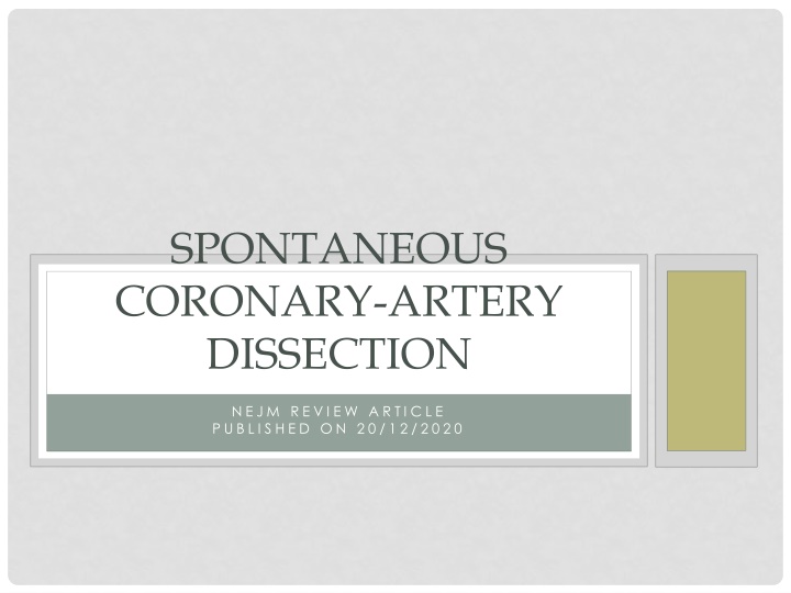
SPONTANEOUS CORONARY-ARTERY DISSECTION N E J M R E V I E W A R T I C L E P U B L I S H E D O N 2 0 / 1 2 / 2 0 2 0
INTRODUCTION Important cause of myocardial infarction in young persons Occurs primarily in women Prevalence of CV risk factors among women with MI from SCAD is lower Accurate and rapid diagnosis is paramount Can be a forme fruste of an underlying systemic arteriopathy namely, fibromuscular dysplasia
DEFINITION Separation of the layers of an epicardial coronary-artery wall by intramural hemorrhage, with or without an intimal tear Not associated with atherosclerosis, iatrogenic injury, or trauma
PATHOPHYSIOLOGY Hypothesis - an intimal tear results in the creation and propagation of a false lumen within the medial layer The presence of a coronary intramural hematoma without evidence of an intimal tear was detected almost 20 years ago with the use of IVUS
Accepted hypothesis - primary event may be medial dissection or rupture of the vasa vasorum resulting in a secondary intramural hemorrhage and formation of an intramural hematoma The final common pathway for the development of acute MI is coronary obstruction due to luminal compression, either by a dissection flap or by propagation of an intramural hematoma
A normal coronary artery appears as a smooth vessel free of luminal irregularities. Asterisks indicate a guidewire shadow artifact. Type 1 spontaneous coronary-artery dissection (SCAD) has the pathognomonic angiographic appearance of an arterial dissection, including multiple radiolucent lumens due to an intimal tear that causes contrast dye to penetrate through two flow channels. A radiolucent flap separating the two flow channels is visible on angiography (left image, arrow). Type 1 SCAD also can result in retention of contrast dye within the intimal tear or slow clearing of contrast material. On optical coherence tomography (OCT), an intimal tear (right image, arrow) separating the true lumen from the false lumen (FL) is shown. Double-headed arrows indicate an intramural hematoma.
Type 2 SCAD is the most common type; 60 to 75% of patients with SCAD have this angiographic diagnosis. Type 2 SCAD is characterized by the absence of an intimal tear and appears as a long segment of diffusely narrowed artery because of an intramural hematoma that causes stenosis of varying severity. Type 2 SCAD lesions are long (typically >20 mm), often appear as an abrupt caliber change in the artery (angiographic images, square brackets), and will either be flanked by an artery of normal caliber (type 2A) or continue to the tip of the artery (type 2B). OCT imaging shows an intramural hematoma. The type 3 variant is the least common type of SCAD and the most challenging type to recognize on angiography. The appearance also suggests compression by an intramural hematoma, but type 3 SCAD is usually 20 mm or less in length. Type 3 SCAD is described as an atherosclerosis mimic, and intracoronary imaging is often necessary to confirm the diagnosis. This type should be considered when there is a high index of suspicion for SCAD, atherosclerosis is absent in the remainder of the coronary vasculature, the lesion is linear and long (11 to 20 mm), or coronary tortuosity is present. As with type 2 SCAD lesions, OCT imaging reveals a compressive intramural hematoma. Intracoronary nitroglycerin can be administered during angiography, particularly when type 2 or type 3 SCAD is suspected, to rule out the possibility that coronary vasospasm is causing the angiographic abnormality
Finally, type 4 SCAD has been described as a complete occlusion of the vessel (arrow). Its appearance may be similar to that of thromboembolic occlusion, and dissection as the cause of occlusion may be evident only when the underlying vessel architecture appears after vessel recanalization or after exclusion of an embolic cause and repeat coronary angiography shows healing of the vessel. Repeat angiography that shows artery healing or the presence of extracoronary arterial findings provides support for the diagnosis of SCAD
EPIDEMIOLOGY Affects both sexes across the life span. 90% are women who present between 47 and 53 years of age The prevalence of typical CV risk factors is lower than among those who have a MI from atherosclerotic disease Cohort studies incorporating direct angiographic review indicate that one quarter to one third of MI in women younger than 50 years of age are caused by SCAD SCAD accounts for approx. 15 to 20% of MI during pregnancy or the peripartum period
ETIOLOGY Cause of SCAD is unknown The commonest identified factors were postpartum, FMD, connective tissue disease and hormonal therapy Factors related to patient vulnerabilities and inciting triggers such as emotional stress, physical stress (e.g., from an extreme Valsalva maneuver, retching, vomiting, coughing, or isometric exercise), the use of stimulant medications or illicit drugs, and hormonal triggers (e.g., pregnancy) Triggers of SCAD shear stress on the coronary artery wall, mediated by elevated catecholamine levels and intra- abdominal pressure
Prevalence of this condition among men is low Men with SCAD have a lower prevalence of anxiety and depression. More often cite a physical stressor (e.g., exercise or heavy lifting) rather than an emotional stressor before symptom onset Tend to have a lower prevalence of FMD than women with SCAD
The predilection of SCAD to affect women disproportionately provides compelling fodder for a hypothesis of a role of hormones in this condition But the percentage of postmenopausal women in SCAD registries is consistently approximately 55%, and the prevalences of nulliparity and multiparity are similar among patients with SCAD.
Among patients with SCAD who are referred for panel based sequencing of genes known to cause aortopathies or connective-tissue diseases (e.g.,vascular Ehlers Danlos syndrome, Marfan s syndrome, and the Loeys Dietz syndrome), the prevalence of a disease-causing mutation is approximately 5 to 8%
There was a high frequency of variants associated with the Loeys Dietz syndrome This finding suggests a role for dysregulated transforming growth factor signaling in the pathogenesis of SCAD None of the patients in that study had clinical features that were typical of the Loeys Dietz syndrome
Currently, apart from genetic screening of first-degree family members of patients with SCAD in whom a monogenic vascular disease has been diagnosed, no firm recommendations are in place for routine genetic or clinical screening of asymptomatic relatives of patients after a SCAD event.
PHACTR1EDN1, a genetic locus associated with migraine headache, cervical artery dissection, and FMD - increased risk of SCAD. Susceptibility genes - LRP1, LINC00310, FBN1, and ADAMTSL4 that are expressed in arteries identified in familial and sporadic SCAD
A polygenic risk score for SCAD derived from genomewide association testing was associated with a decreased risk of atherosclerotic coronary artery disease Genetic studies formulate the basis for a thesis that myocardial infarction due to SCAD is a pathophysiological entity that is distinct from myocardial infarction due to atherosclerosis.
A consistent finding in registries of patients with SCAD is the high prevalence of concomitant noncoronary arterial abnormalities More than 50% of the patients also had FMD Autopsy and intracoronary imaging studies suggest that SCAD may be an initial manifestation of FMD
Abnormalities known to be associated with FMD are cervical- artery, visceral-artery, and peripheral-artery aneurysm, as well as pseudoaneurysm, dissection, and tortuosity, but these findings have also been reported in multiple cohorts of patients with SCAD, even in the absence of diagnosed concurrent FMD Cerebral aneurysm - detected in 7 to 14% of patients with SCAD who have undergone screening
Whether or not SCAD is a coronary manifestation of FMD, it is clear that SCAD may be a forme fruste of an underlying systemic arteriopathy that leaves the affected patient vulnerable to dissection when exposed to arterial shear stress related to an inciting trigger
EXAMPLES A 70-year-old woman with an acute MI presented with VF and SCAD of an obtuse marginal branch of the left circumflex artery. Computed tomographic angiography (CTA) of the abdomen and pelvis revealed a celiac-artery dissection and multifocal FMD of the left external iliac artery
A 57-year-old woman presented with NSTEMI and SCAD of the obtuse marginal branch of the left circumflex artery. Limited femoral angiography of the right external iliac artery at the completion of coronary angiography revealed mild aneurysmal changes and beading that were consistent with multifocal FMD. CTA also showed FMD of the right renal artery
A 35-year-old woman had SCAD of the left anterior descending artery 2 weeks after delivering her fourth child. She had had a left internal carotid artery dissection 10 years before SCAD, and CTA of the head and neck showed a dissection of the left internal carotid artery and multifocal FMD of the right internal carotid artery and bilateral vertebral arteries.
CLINICAL SIGNS AND SYMPTOMS In more than 90% of patients who survive to initial evaluation, SCAD manifests as MI 20 to 50% of patients present with STEMI, 3 to 5% present with ventricular arrhythmias for which cardioversion is warranted and 2% present in cardiogenic shock Chest pain, the chief symptom reported in 85 to 96% of patients, is variably associated with radiation of pain to the arm, neck, or back; dyspnea; and diaphoresis
DIAGNOSIS SCAD is diagnosed with angiography. Patients in whom SCAD is the suspected cause of acute MI should undergo coronary angiography: to confirm the diagnosis define high-risk anatomical features that would warrant consideration of early revascularization Early recognition is critical because there are important differences between the management of SCAD and the management of atherosclerotic acute MI
SCAD is classified primarily differentiated by the presence or absence of an intimal tear SCAD can occur in any coronary artery LAD and its branches are most commonly involved Multivessel SCAD involving noncontiguous arteries occurs in approximately 10 to 15% of patients with SCAD Majority of SCAD lesions can be diagnosed with angiography alone, it may be difficult to distinguish a potential case of SCAD from other causes of coronary-artery stenosis.
The favored approach for patients with SCAD who are in clinically stable condition is medical treatment When angiography is not diagnostic for SCAD, intravascular ultrasonography or OCT may be used for confirmation Angiography provides a lumenogram of the artery, intravascular imaging particularly OCT, with its high axial spatial resolution (15 m) can confirm the diagnosis of SCAD by showing the true and false lumens, intramural hematoma, dissection flaps, fenestrations, and entry tears connecting true and false lumens
Intravascular imaging complications: 1. Extension of the dissection, 2. Impaired flow after acquisition of intravascular imaging, 3. Iatrogenic dissection, and 4. Cannulation of the false lumen Intracoronary imaging is reserved for situations in which angiography is not diagnostic for SCAD or when intracoronary imaging is used for guidance during PCI
Coronary computed tomographic angiography (CCTA) can be used to visualize dissection flaps, stenoses, and intramural hematomas Useful particularly in cases of proximal lesions Limitation: Noncalcified atherosclerotic plaque can be mistaken for an intramural hematoma, and the spatial resolution of CCTA for small vessels limits visualization of the distal portion of the vessels that is often affected by SCAD
In some situations, SCAD is strongly suspected but angiographic findings are uncertain ancillary imaging techniques are not available or not diagnostic. Clinical features to support a SCAD diagnosis include coronary tortuosity on angiography, the presence of FMD in another arterial bed, and a reduction in arterial stenosis (as evidence of vessel healing) on repeat angiography.
MANAGEMENT OF ACUTE MI Acute MI caused by SCAD has three major differences with atherosclerotic coronary lesions 1. The underlying pathophysiological feature of SCAD is medial dissection, not plaque rupture or erosion in an inflammatory and thrombotic milieu 2. PCI for SCAD is challenging and is associated with worse short- and long term outcomes than those associated with PCI for atherosclerotic lesions 3. The majority of medically treated SCAD lesions show angiographic evidence of healing over time, with restoration of blood flow and a decrease in the severity of stenosis
More than 80% of patients can be successfully treated medically Expert consensus suggests that medical management is preferred over immediate revascularization Recurrent SCAD (a new SCAD event that is temporally separated from the index event) tends to occur in different vessels from those in the initial dissection, revascularization has not been shown in long-term follow-up to prevent recurrent myocardial infarction due to SCAD
MEDICAL THERAPY VS REVASCULARIZATION Factors to consider include the patient s clinical status, the territory at risk, the amount of myocardium at risk, and the degree of distal flow in the affected vessel. High-risk clinical features include persistent chest pain with evidence of ongoing or worsening ischemia, hemodynamic instability, shock, or clinically significant ventricular arrhythmias High-risk anatomical features include involvement of multivessel severe proximal dissections or of the left main artery or the ostial left anterior descending artery
Experts advocate avoiding revascularization attempts if the patient is in stable condition and has adequate distal flow (TIMI grade 2 or 3) in the vessel, even if there is clinically significant stenosis When high-risk features are present, consideration of immediate revascularization is warranted, with the decision to perform PCI or refer the patient for coronary-artery bypass grafting (CABG)
PCI IN SCAD Long lesions may warrant the use of multiple stents, coronary wires can enter the false lumen and cause vessel occlusion, tortuous coronary arteries may be prone to iatrogenic injury, and hematoma propagation can result in the loss of distal- vessel patency or retrograde extension to more proximal vessels Possible late term adverse events - stent malapposition as a result of resorption of intramural hematoma over time These factors contribute to PCI success rates that range from 47% to 72% in large cohort studies
CABG IN SCAD High-risk anatomical lesions (e.g., left main and multiple proximal dissections) Attempts at PCI have failed PCI is thought to be of prohibitive risk Large areas of myocardium at risk Medical therapy alone is not sufficient to treat ongoing ischemia
Short-term success with CABG is high Long-term patency of bypass grafts is poor Recanalization of the native coronary arteries results in competitive flow and subsequent graft occlusion. Experts recommend the use of vein grafts to revascularize SCAD in an effort to conserve potential arterial conduits for later use
The incidence of inhospital recurrent myocardial infarction or unplanned revascularization is 5 to 10%, and among patients who have received medical treatment, the risk of angiographically confirmed dissection extension is as high as 17% over a period of 14 days Coronary angiography should be considered for determination of the need for revascularization in patients with clinical deterioration.
Among patients with acute myocardial infarction due to SCAD who are readmitted within 30 days after hospital discharge, 45% present with recurrent acute myocardial infarction, of whom half present within 2 days after discharge. Revascularization may improve flow within the dissected vessel, it does not protect against the risk of dissection extension, and PCI may result in an increased incidence of hospital readmission at 30 days For these reasons, the current consensus is that hospitalization for 3 to 5 days in patients with an acute myocardial infarction due to SCAD is reasonable in order to observe for adverse ischemic events
ANTICOAGULATION Anticoagulation and dual antiplatelet therapy are often initiated before SCAD is diagnosed. The treatment of any existing luminal thrombosis is balanced against the hypothetical risk of dissection extension due to worsening of intramural bleeding. In the absence of clear alternative indications anticoagulation should be discontinued after SCAD has been confirmed on angiography The use of thrombolysis for the management of acute SCAD is not recommended
ANTIPLATELET THERAPY Approximately 90% of patients who have received a diagnosis of SCAD are discharged with at least one antiplatelet agent The expert consensus is that dual antiplatelet therapy may be considered during the acute phase of SCAD and for up to 1 year for patients who receive medical treatment The duration of antiplatelet therapy should be determined for each patient. For instance, aspirin may be considered in patients with FMD to prevent thrombotic and thromboembolic complications, whereas a shorter duration of therapy may be warranted in premenopausal women with excess bleeding from menorrhagia who do not have other indications for antiplatelet therapy
BETA-BLOCKERS, ARBS AND ACEI Major societal guidelines for the use of beta-blockers, ARBs, and ACEI in the treatment of acute myocardial infarction and heart failure should be followed for the use of these agents in patients with SCAD Beta-blockers may have an additional benefit of preventing the recurrence of SCAD In a single-center observational study involving 327 patients, the use of beta-blockers was associated with a 64% decrease in the incidence of recurrent SCAD over a median of 3.1 years.
STATINS SCAD is not mediated by atherosclerotic plaque rupture Data are lacking to support the routine use of statins after myocardial infarction due to SCAD
ANTIANGINAL THERAPY Accounts for 20% of readmissions within 30 days after acute myocardial infarction due to SCAD Chest pain in patients with abnormal ischemia testing should be treated with medical therapy and investigated with further cardiac testing Coronary vasospasm, endothelial dysfunction, microvascular disease, catamenial chest pain, and noncardiac chest pain should be considered in patients who continue to have atypical chest pain that is not associated with abnormal ischemia testing Nitrates, calcium-channel blockers, and ranolazine are potential therapies to consider
PREVENTION OF SCAD RECURRENCE Mortality after SCAD is low 1% over a period of 3 years The incidence of recurrent myocardial infarction is substantial, with 17 to 18% of patients having recurrent myocardial infarction over a span of 3 to 4 years The majority of these recurrent myocardial infarctions are due to recurrent SCAD


 can")