Staphylococcus aureus Characteristics and Pathogenesis
Explore the features of Staphylococcus aureus, including its Gram-positive nature, cell wall factors, toxins, virulence factors, enzymes, pathogenesis of infection, and clinical manifestations such as skin and soft tissue infections.

Download Presentation

Please find below an Image/Link to download the presentation.
The content on the website is provided AS IS for your information and personal use only. It may not be sold, licensed, or shared on other websites without obtaining consent from the author. If you encounter any issues during the download, it is possible that the publisher has removed the file from their server.
You are allowed to download the files provided on this website for personal or commercial use, subject to the condition that they are used lawfully. All files are the property of their respective owners.
The content on the website is provided AS IS for your information and personal use only. It may not be sold, licensed, or shared on other websites without obtaining consent from the author.
E N D
Presentation Transcript
General Features Gram positive cocci in grape-like clusters Catalase positive
Cell wall associated factors Cell wall associated factors Peptidoglycan Rigidity, Endotoxin Teichoic acid Adhesion, Inhibit opsonisation Cell surface adhesins-Clumping factor Protein A Adhesion, Clumping Anti-complementary Chemotactic, Mitogenic, Inhibit opsonisation Induction of platelet damage IgG Fc Co-agglutination Capsule Inhibit phagocytosis
Toxins Toxins Membrane active toxins Hemolysins- , , , Leucocidin F & S toxins + hemolysin and leucocidal (Synergohymenotropy) hemolytic Epidermolytic toxin Staphylococcal scalded-skin syndrome Enterotoxins Serotype A- MC, Superantigen Self limiting food poisoning IP<6 Hrs Preformed toxin vagus stimulation Toxic shock syndrome toxin Super antigen Risk - abscesses, osteomyelitis, post- surgical wound infection
Virulence Factors hemolysin Inactivated at 70 C reactivated at 100 C Lethal, leucocidal, dermonecrotic, cytotoxic and neurotoxic Lyses rabbit RBCs, but less active against sheep and human RBCs. Sphingomyelinase Lyses sheep RBC, but not human or rabbit RBC Exhibits hot-cold phenomenon Act together with leucocidin for hemolytic activity. Lyses rabbit, sheep and human RBCs Surfactant action Lyses rabbit, sheep, horse & human RBCs Lethal, leucocidal and dermonecrotic hemolysin hemolysin hemolysin
Enzymes Extracellular Enzymes Coagulase + CRF Tube coagulase test prothrombin to thrombin Heat stable thermonuclease specific to S.aureus Deoxyribonuclease specific to S.aureus Lipase Protease Staphylokinase(fibrinolysin) Hyaluronidase
Pathogenesis Colonization-anterior nares, axilla and perineal skin Introduction into the tissue- minor abrasions or instrumentation adhere to tissue Invasion- with help of enzymes Evasion of host defence mechanisms - Anti-phagocytic -microcapsule and Protein A - Inhibition of leukocyte migration - -formation of small colony variants Metastatic spread- hematogenous
Clinical Manifestations Skin & Soft tissue infections Folliculitis, Furuncle Carbuncle, Impetigo Mastitis and breast abscess Surgical site wound infections Cellulitis Hidradenitis suppurativa
Clinical Manifestations... Musculoskeletal Infections Septic arthritis Osteomyelitis Pyomyositis in HIV Psoas abscess Epidural abscess Respiratory Tract Infections Ventilator associated pneumonia in adults Septic pulmonary emboli Post viral pneumonia Empyema and Pneumothorax Pneumatocele in neonates
Bacteremia and its Complications Bacteremia and its Complications Sepsis, septic shock Central line associated blood stream infection Metastasis - kidney, joints, bone and lung Native-valve endocarditis Prosthetic-valve endocarditis Intravenous drug use associated endocarditis
Clinical Manifestations Clinical Manifestations Toxin-Mediated Illnesses Toxic shock syndrome Food poisoning Staphylococcal scalded- skin syndrome Infections Associated with MRSA Necrotizing pneumonia Necrotizing fasciitis
Staphylococcal Gastroenteritis Staphylococcal Gastroenteritis Enterotoxin - expressed by 50% of S.aureus preformed heat stable toxin and resistant to gastric juice Serotypes(A E, G I, R-T and V). Type A Most common Site of action- Stimulates the vagus nerve and the vomiting center. Stimulation of intestinal peristaltic activity
Staphylococcal Gastroenteritis Staphylococcal Gastroenteritis incubation period 16 Hrs Symptom- nausea, vomiting and occasionally diarrhea, hypotension, and dehydration. No fever. Selflimiting within 8 10 hour. Source - food handler Food items - milk products, bakery food,custards, potato salad, or processed meats.
Staphylococcal scalded Staphylococcal scalded- -skin syndrome (SSSS) skin syndrome (SSSS) Epidermolytic/exfoliative toxin Two proteins- ET-A (chromosomal and heat stable), ET- B (plasmid coded, heat labile). More common in newborns and children - May vary from localized tender blisters& bullae formation to exfoliation & separation of outer epidermal layer leaving denuded underlying skin (Nikolsky's sign).
Staphylococcal scalded Staphylococcal scalded- -skin syndrome (SSSS) skin syndrome (SSSS) Milder form - pemphigus neonatorum and bullous impetigo Ritter s syndrome - Severe form in newborns - Fever, lethargy, and irritability with poor feeding. - S.aureus bacteriophage group II
TOXIC SHOCK SYNDROME TOXIC SHOCK SYNDROME Toxic shock syndrome toxin (TSST)- Enterotoxin F, a superantigen Risk factors- vaginal tampons, abscesses, osteomyelitis and post- surgical wound infection. Pathogenesis- TSST absorbed into circulation - non-specific T cells stimulation - excessive cytokine production -multisystem disease.
TOXIC SHOCK SYNDROME TOXIC SHOCK SYNDROME Clinical features- Fever, hypotension, mucosal hyperemia, vomiting, diarrhea, confusion, myalgia, abdominal pain and erythematous rashes rapid involvement of liver, kidneys, GIT and/or CNS. Diagnosis- - Detection of TSST - latex agglutination test and ELISA - TSST genes 1 and 2 - PCR Treatment- Clindamycin (MSSA), vancomycin (MRSA)
LABORATORY DIAGNOSIS.. LABORATORY DIAGNOSIS.. Infection Suppurative lesion Respiratory Infections Urinary tract infection PUO, Bacteremia Food poisoning Carriers Specimen Pus, wound swab Sputum Mid stream urine Blood Feces, vomitus, food Nasal & perianal swab
LABORATORY DIAGNOSIS.. LABORATORY DIAGNOSIS.. MICROSCOPY Gram staining -Gram positive cocci in clusters
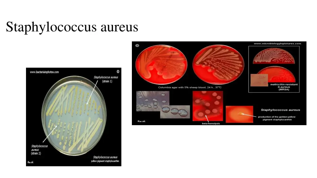
LABORATORY DIAGNOSIS LABORATORY DIAGNOSIS- - CULTURE CULTURE Aerobe, facultative anaerobe, Non-fastdious Nutrient agar Golden yellow Blood agar beta hemolytic
Coagulase test LABORATORY DIAGNOSIS LABORATORY DIAGNOSIS- - Biochemical Tests o Tube coagulase o Clumping factor Heat stable thermo nuclease test Biochemical Tests DNase test Phosphatase (also produced by S.epidermidis) S.aureus positive and CoNS mostly negative Golden yellow pigmentation Hemolysis on blood agar Mannitol fermentation Black coloured colonies on potassiumtellurite agar Gelatin liquefaction Protein A detection
Typing of Typing of S.aureus S.aureus Phenotypic methods - Bacteriophage typing - Antibiogram typing. Genotypic methods - PCR-RFLP
CONTROL MEASURES CONTROL MEASURES Proper hand washing Screening of MRSA carriers Treatment of carriers Stoppage of antibiotic misuse
References Textbook of Medical Microbiology by Ananthnarayan and Paniker Textbook of Medical Microbiology by D.R Arora