Structure and Function of the Immune System
The immune system comprises lymphoid organs, lymphoid cells, other immune cells, and cytokines. Central lymphoid organs like the thymus and bone marrow play crucial roles in hematopoiesis and immune cell development. Lymphoid cells, including T cells, B cells, and NK cells, are vital for immune responses. Phagocytes, dendritic cells, and mast cells are also key components. Cytokines play a pivotal role in immune signaling. Understanding the structure and function of these components is essential for comprehending immune system processes.

Download Presentation

Please find below an Image/Link to download the presentation.
The content on the website is provided AS IS for your information and personal use only. It may not be sold, licensed, or shared on other websites without obtaining consent from the author.If you encounter any issues during the download, it is possible that the publisher has removed the file from their server.
You are allowed to download the files provided on this website for personal or commercial use, subject to the condition that they are used lawfully. All files are the property of their respective owners.
The content on the website is provided AS IS for your information and personal use only. It may not be sold, licensed, or shared on other websites without obtaining consent from the author.
E N D
Presentation Transcript
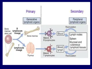
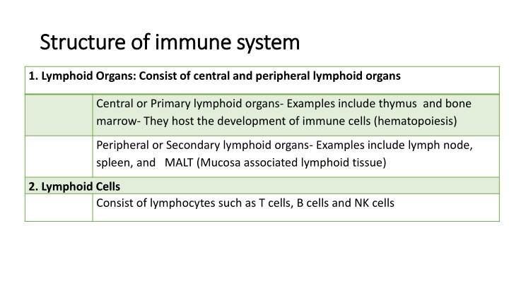
Structure of immune system Structure of immune system 1. Lymphoid Organs: Consist of central and peripheral lymphoid organs Central or Primary lymphoid organs- Examples include thymus and bone marrow- They host the development of immune cells (hematopoiesis) Peripheral or Secondary lymphoid organs- Examples include lymph node, spleen, and MALT (Mucosa associated lymphoid tissue) 2. Lymphoid Cells Consist of lymphocytes such as T cells, B cells and NK cells
Structure of immune system Structure of immune system 3.Other cells of immune system- Include phagocytes such as macrophage and microphages (neutrophil, eosinophil & basophil), dendritic cells, mast cells and platelets. 4.Cytokines- They are the soluble products secreted from various cells of immune system Include interleukins, interferons, tumor necrosis factors, colony stimulating factors, etc.
Central lymphoid organ Central lymphoid organ - - Bone marrow Bone marrow All the cells in blood are originated from pluripotent hematopoietic stem cells of bone marrow and the process is called a hematopoiesis. In early fetal life, hematopoiesis occurs in liver; gradually the stem cells migrate to bone marrow. By birth, the stem cells occupy most of the bone marrow space of large bones.
Central lymphoid organ Central lymphoid organ - - Bone marrow As the individual ages, hematopoietic activity in large bones decreases and after puberty hematopoiesis is mostly confined to axial bones such as pelvis, vertebrae, sternum, skull & ribs. Progenitor T and B cells originate in bone marrow. oFurther development of B cells occurs in bone marrow itself. oProgenitor T cells migrate to thymus for further proliferation. Bone marrow
Central lymphoid organ Central lymphoid organ - - Thymus Site of proliferation and maturation of T cells. Development: o Developed in the embryonic life (third month) from third/fourth pharyngeal pouch. oHighly active at birth, continues to grow for many years, reaches its peak size at puberty, and then it degenerates. Structure: Thymus has two lobes surrounded by a fibrous capsule. Septa arising from capsule divide thymus into lobules, and each lobule is differentiated into an outer cortex & an inner medulla Thymus
Thymus Thymus - -Cortex Densely populated and contains: oThymocytes- Lymphocytes of thymus (immature and many in number). oCortical epithelial cells and oNurse cells (specialized epithelial cells with long membrane extensions that surround many thymocytes) Cortex
Thymus Thymus - -Medulla Sparsely populated and contains: oThymocytes which are relatively more mature and fewer in number oMedullary epithelial cells oInterdigitating dendritic cells oHassall s corpuscles (concentric layers of degenerating epithelial cells) Medulla
Thymic Thymic hormones hormones Produced from the epithelial cells of thymus. They are believed to attract the precursor T cells (progenitor T cells) from bone marrow. oThymulin oThymopoietin oThymosin
Maturation of T cells Maturation of T cells Cell to cell interaction between thymocytes & thymic stromal cells (including epithelial cells, dendritic cells & macrophages) Effect of thymic hormones.
Central tolerance Central tolerance Only 2-5% of the developing T cells become mature and released out from thymus. Remaining T cells are destroyed as they are either not capable of recognizing MHC or are believed to be self-reacting in nature. Destruction of such self-reacting T cells prevents development of autoimmunity (immune response against self-antigens). Such tolerance to self-antigens mediated by thymus that occurs in embryonic life is called as central tolerance.
Defect in thymus Defect in thymus Leads to defect in maturation of T lymphocytes that in turn results in severe life threatening cell mediated immunodeficiency disorders. DiGeorge syndrome (immunodeficiency disorder) - congenital aplasia of thymus. Nude mice- Mice with congenital absence of thymus.
PERIPHERAL LYMPHOID ORGAN PERIPHERAL LYMPHOID ORGAN - - Lymph Node Lymph Node Small bean shaped organs. Occur in clusters or in chains, distributed along the length of lymphatic vessels. Lymph nodes act as physiological barriers - filter the microbial antigens carried to lymph node by activating the T and B cells.
Lymph Node Lymph Node - - Structure Thymus is divided into three parts: oCortex oMedulla (both are B cell areas) oParacortex (T cell area). Bears the lymphatic vessels (efferent and afferent) and blood vessels. Structure
Lymph Node Lymph Node - - Structure Thymus is divided into three parts: oCortex oMedulla (both are B cell areas) oParacortex (T cell area) Bears the lymphatic vessels (efferent and afferent) and blood vessels. Structure
Lymph Node Lymph Node - - Cortex Contains lymphoid follicles that are composed of: oB cells oFew special type of dendritic cells (called follicular dendritic cells) oOccasional T cells. Lymphoid follicles are mainly of two types: oPrimary lymphoid follicles oSecondary lymphoid follicles Cortex
Primary lymphoid follicles Primary lymphoid follicles Found before the antigenic stimulus. Smaller Contain resting B cells.
Secondary lymphoid follicles Secondary lymphoid follicles Following contact with an antigen - resting B cells starts dividing and become activated. Activated B cells differentiate rapidly in to plasma cells and memory B cells. Follicles become larger called secondary lymphoid follicles.
Secondary lymphoid follicles Secondary lymphoid follicles Has two areas: oCentral area - germinal center oPeripheral zone - mantle area; contains activated B cells.
Lymph Node Lymph Node Paracortical Paracortical area and Medulla area and Medulla Paracortical are: oPresent in between cortex and medulla. oT cell area of lymph node (rich in naive T cells). oContains macrophages and interdigitating dendritic cells, which trap the antigens and present to T cells. Medulla: oInnermost area of lymph node. oRich in B-lymphocytes (mainly plasma cells).
Spleen Spleen Largest secondary lymphoid organ. Acts as physiological barrier similar to lymph node in clearing the microbial antigens through the antigenic stimulation of T and B cells.
Spleen Spleen - - Structure Structure Situated below the diaphragm on left side of the abdomen. Adult spleen measures about 5-inch in length & weighs around 150gm. Divided into two compartments: central white pulp and outer red pulp, surrounded by capsule and intervened by trabeculae.
Spleen Spleen - - Structure Structure White pulp: oCentral densely populated area. oContains T cells and B cells. oIt has two parts: Periarteriolar lymphoid sheath (PALS), which is T cell area Marginal zone is located peripheral to the PALS and is populated by B cell lymphoid follicles
Spleen Spleen - - Structure Structure Red pulp: oArea that surrounds the sinusoids. oFilled with red blood cells (RBCs). oOlder and defective RBCs are destroyed here.
Defect in spleen Defect in spleen Increased incidence of bacterial sepsis caused primarily by capsulated bacteria such as: oStreptococcus pneumoniae oNeisseria meningitides oHaemophilus influenzae.
Refrences Textbook of Medical Microbiology by Ananthnarayan, Paniker Textbook of Medical Microbiology by Satish Gupte Textbook of Medical Microbiology by S. Bhat, A.S.Sastry Textbook of Medical Microbiology by D.R.Arora, Brij bala Arora