Structure of the Helix: Four Models Displayed
Explore four different models showcasing the structure of the helix, from the formation of a right-handed helix to the helix dipole and the conformation of polypeptide chains. Gain insights into its intricacies and visual representations through these informative images.

Download Presentation

Please find below an Image/Link to download the presentation.
The content on the website is provided AS IS for your information and personal use only. It may not be sold, licensed, or shared on other websites without obtaining consent from the author.If you encounter any issues during the download, it is possible that the publisher has removed the file from their server.
You are allowed to download the files provided on this website for personal or commercial use, subject to the condition that they are used lawfully. All files are the property of their respective owners.
The content on the website is provided AS IS for your information and personal use only. It may not be sold, licensed, or shared on other websites without obtaining consent from the author.
E N D
Presentation Transcript
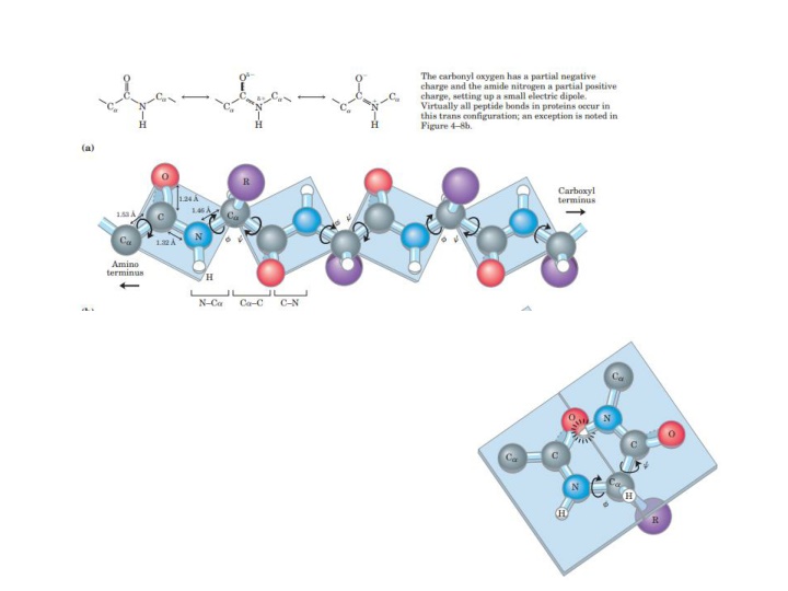
Four models of the helix, showing different aspects of its structure. (a) Formation of a right-handed helix. The planes of the rigid peptide bonds are parallel to the long axis of the helix, depicted here as a vertical rod. (b) Ball- and-stick model of a right handed helix, showing the intrachain hydrogen bonds. The repeat unit is a single turn of the helix, 3.6 residues. (c) The helix as viewed from one end, looking down the longitudinal axis (derived from PDB ID 4TNC). Note the positions of the R groups, represented by purple spheres. This ball-and-stick model, used to emphasize the helical arrangement, gives the false impression that the helix is hollow, because the balls do not represent the van der Waals radii of the individual atoms. As the space-filling model (d) shows, the atoms in the center of the helix are in very close contact