Study of Structural Diseases in Pathology: Rudolf Virchow's Cellular Concept
Pathology is the study of structural diseases affecting organs and systems. In the 19th century, Rudolf Virchow proposed that injury to the smallest living cell must maintain a functional barrier between its internal systems. The plasma membrane plays a crucial role in maintaining the cell's internal environment and house signaling molecules for communication. Pathology explores how cells adapt to stressors, with persistent stress leading to chronic cell injury. Understanding cellular responses to injury, such as atrophy, hypertrophy, and neoplasia, is essential in studying disease pathology and adaptive mechanisms. The concept of proteasomes in regulating cellular homeostasis and response to stress highlights the importance of selective protein elimination pathways.

Download Presentation

Please find below an Image/Link to download the presentation.
The content on the website is provided AS IS for your information and personal use only. It may not be sold, licensed, or shared on other websites without obtaining consent from the author.If you encounter any issues during the download, it is possible that the publisher has removed the file from their server.
You are allowed to download the files provided on this website for personal or commercial use, subject to the condition that they are used lawfully. All files are the property of their respective owners.
The content on the website is provided AS IS for your information and personal use only. It may not be sold, licensed, or shared on other websites without obtaining consent from the author.
E N D
Presentation Transcript
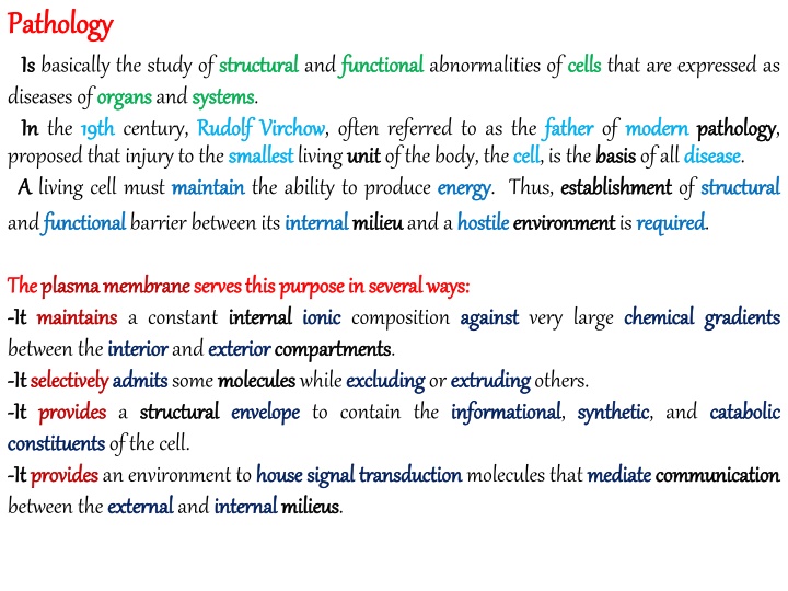
Pathology Pathology Is Is basically the study of structural diseases of organs organs and systems In In the 19 19th th century, Rudolf proposed that injury to the smallest A A living cell must maintain and functional functional barrier between its internal structural and functional systems. Rudolf Virchow Virchow, often referred to as the father smallest living unit unit of the body, the cell maintain the ability to produce energy internal milieu milieu and a hostile functional abnormalities of cells cells that are expressed as father of modern cell, is the basis basis of all disease energy. Thus, establishment establishment of structural hostile environment environment is required modern pathology pathology, disease. structural required. The The plasma plasma membrane - -It It maintains maintains a constant internal between the interior interior and exterior - -It It selectively selectively admits admits some molecules - -It It provides provides a structural structural envelope constituents constituents of the cell. - -It It provides provides an environment to house between the external external and internal membrane serves serves this internal ionic exterior compartments compartments. molecules while excluding envelope to contain the informational this purpose purpose in in several ionic composition against several ways ways:: against very large chemical chemical gradients gradients excluding or extruding extruding others. informational, synthetic synthetic, and catabolic catabolic house signal internal milieus milieus. signal transduction transduction molecules that mediate mediate communication communication
conditions (stresses oxygen supply; stresses or or The The cell Injury the presence of noxious Patterns Patterns of response to such stresses exceeds exceeds the adaptive capacity capacity of the cell, the cell dies and the expression expression of a cells preexisting cell must must be able to adapt Injury ), such as as changes in temperature noxious agents; and so so on on. adapt to adverse temperature, solute concentrations adverse environmental conditions concentrations, or oxygen stresses is the cellular basis dies. Thus, pathology preexisting capacity capacity to adapt basis of disease pathology is the study adapt to such injury injury. disease. If If an injury study of cell injury injury Reactions Reactions to to Persistent Persistent Persistent stress often leads to chronic associated with the death death of individual cells By By contrast contrast, the cellular response to persistent sublethal reflects adaptation adaptation of the cell to a hostile The The major major adaptive responses are atrophy, hypertrophy intracellular intracellular storage storage. In In addition addition, certain forms of neoplasia Persistent Stress Stress and and Cell Cell Injury Injury chronic cell cells. cell injury injury. In general, permanent permanent organ injury injury is sublethal injury injury, whether chemical chemical or physical physical, hostile environment. hypertrophy, hyperplasia hyperplasia, metaplasia metaplasia, dysplasia dysplasia, and neoplasia may follow follow adaptive adaptive responses.
Proteasomes Proteasomes are Altered Altered Extracellular Extracellular Environment Cellular homeostasis homeostasis requires mechanisms selectively selectively. Although there is evidence that more understood mechanism by which cells target specific proteins for elimination ( (Ub Ub) )- -proteasomal proteasomal apparatus apparatus. are Key Key Participants Participants in in Cell Environment Cell Homeostasis Homeostasis,, Response Response to to Stress Stress,, and and Adaptation Adaptation to to mechanisms that allow the cell to destroy more than than one destroy certain proteins exist, the best elimination is the ubiquitin proteins best ubiquitin one such pathway may exist Ubiquitin Ubiquitin and Proteins Proteins to be degraded Ub is a 76 76- -amino amino acid selective protein elimination destroyed destroyed. The process of attaching Ub to proteins is called and Ubiquitination Ubiquitination degraded are flagged acid protein that is almost identical in yeast elimination: it is conjugated to proteins as a flag flagged by attaching small chains of ubiquitin ubiquitin molecules to them. yeast as in humans humans. It is the key flag to identify identify those proteins to be called ubiquitination ubiquitination. key to
Ubiquitin Ubiquitin- -proteasome proteasome pathways by which ubiquitin (Ub) targets proteins for specific elimination elimination in proteasomes are shown here. Ub is activated (Ub Ub* *) by E E1 1 ubiquitin enzyme enzyme, then transferred to an E E2 2 (ubiquitin conjugating conjugating enzyme enzyme). The E E2 2- -Ub* interacts with an E3 (ubiquitin ubiquitin ligase particular particular protein protein. The process may be repeated multiple times to append a chain of Ub These complexes may be deubiquitinated ubiquitinating enzymes (DUBs DUBs). If degradation is to proceed, 26 26S S proteasomes proteasomes recognize the poly- Ub-conjugated protein via their 19 19S S subunit degrade degrade it into oligopeptides oligopeptides. In the process, Ub moieties are returned to the cell pool monomers monomers. pathways.. The mechanisms ubiquitin activating activating ubiquitin Ub* complex ligase) to bind a Ub moieties moieties. deubiquitinated by de- subunit and pool of ubiquitin
Atrophy Atrophy Is Is an an Adaptation - -Clinically Clinically, atrophy atrophy is often noted as decreased - -It It either pathologic pathologic or physiologic Thus, atrophy may result from disuse of normal aging aging. -One One must distinguish distinguish the organ Reduction Reduction in an organ's organ's size example example, atrophy of the brain of the organ cannot cannot be restored Adaptation to to Diminished Diminished Need decreased size Need or or Resources Resources for for a a Cell's size or function function of an organ. Cell's Activities Activities physiologic. disuse of skeletal muscle or or from loss loss of of trophic trophic signals as part organ atrophy from size may reflect reversible brain in Alzheimer Alzheimer disease is secondary to extensive cell restored . from cellular cellular atrophy. reversible cell atrophy or irreversible irreversible loss of cells. For cell death death; the size Causes Causes of of atrophy 1 1- -Reduced Reduced Functional treatment for a bone fracture 2 2- -Inadequate Inadequate Supply Supply of of Oxygen -Total Total cessation cessation of oxygen perfusion of tissues results in cell - when oxygen deprivation is insufficient atrophy:: Functional Demand fracture or after prolonged bed rest. Oxygen:: Demand:: For example, after immobilization immobilization of a limb limb in a cast as cell death death. partial ischemia ischemia ), cell atrophy insufficient to kill kill cells (partial atrophy is common.
3 3- -Insufficient Insufficient Nutrients Starvation Starvation or inadequate particularly in skeletal Nutrients inadequate nutrition associated with chronic skeletal muscle muscle. chronic disease leads to cell atrophy atrophy, 4 4- -Interruption Interruption of of Trophic The functions functions of many cells depend on signals transmitted by chemical endocrine endocrine system and neuromuscular neuromuscular transmission are the best hormones hormones or, for skeletal skeletal muscle, synaptic synaptic transmission, place These can be eliminated by removing removing the source gland gland or denervation denervation. If the anterior anterior pituitary is surgically resected adrenocorticotropic hormone (ACTH ACTH, also termed corticotropin hormone (FSH FSH) results in atrophy atrophy of the thyroid Trophic Signals Signals chemical mediators best examples. The actions of place functional functional demands on cells. source of the signal signal e.g., via ablation mediators. The ablation of an endocrine endocrine resected, loss of thyroid-stimulating hormone (TSH corticotropin), and follicle-stimulating thyroid, adrenal adrenal cortex cortex, and ovaries TSH), ovaries, respectively. 5 5- -Persistent Persistent Cell Persistent Persistent cell injury is most commonly caused by chronic prolonged viral viral or bacterial bacterial infections, immunologic example example is the atrophy atrophy of the gastric gastric mucosa Cell Injury Injury chronic inflammation inflammation associated with immunologic and granulomatous granulomatous disorders. A good mucosa that occurs in association with chronic chronic gastritis gastritis.
Aging Aging One of the hallmarks and heart heart, is cell brain brain is invariably decreased the term senile senile atrophy hallmarks of aging, particularly in nonreplicating cell atrophy atrophy. The size size of all parenchymal decreased, and in the very atrophy has been used. nonreplicating cells such as those of the brain parenchymal organs decreases decreases with age very old old the size size of the heart heart may be so diminished brain age. The size diminished that size of the Atrophy Atrophy involves skeletal skeletal muscle muscle, When the need for contraction selective adaptive adaptive mechanisms mechanisms: 1 1- - Protein Protein synthesis synthesis: Shortly after unloading, protein synthesis decreases 2 2- -Protein Protein degradation degradation. Ubiquitin Ubiquitin- -related related specific protein degradation pathways are activated They lead to decreases in specific contractile contractile proteins proteins. 3 3- -Gene Gene expression expression:: There are selective decreases decreases in transcription things, contractile contractile activities activities. 4 4- -Signaling Signaling:: More More complex complex,, and and less less well well understood understood.. 5 5- - Energy Energy utilization utilization:: A A selective selective decrease decrease in in use use of of free involves changes changes both both in in production production and contraction decreases and destruction destruction of of cellular decreases (unloading unloading), cells institute several cellular constituents, constituents, ex. In decreases. activated. transcription of genes for, among other free fatty fatty acids acids..
Atrophy Atrophy of of the photograph photograph of of the the brain brain.. Marked the brain brain.. The Marked atrophy The gyri gyri are are thinned atrophy of of the thinned and the frontal frontal lobe and the the sulci sulci conspicuously conspicuously widened lobe is is noted noted in in this widened.. this
B A A. Proliferative Proliferative endometrium endometrium.. A A.. A A section age age reveals reveals a a thick thick endometrium endometrium composed stroma stroma.. B B.. The The endometrium endometrium of of a a 75 75- -year magnification) magnification) is is thin thin and and contains section of of the composed of of proliferative year- -old contains only only a a few few atrophic the uterus uterus from proliferative glands old woman woman (shown atrophic and and cystic from a a woman woman of of reproductive glands in in an an abundant (shown at at the cystic glands glands.. reproductive abundant the same same
Hypertrophy Hypertrophy Hypertrophy Hypertrophy Is an Increase in Cell Size and Functional Capacity When trophic trophic signals signals or functional demand lead to increased cellular size size (hypertrophy (hyperplasia hyperplasia). - -In In organs organs made of terminally terminally differentiated responses are accomplished solely solely by increased cell size - -In In other other organs 0f not terminally terminally differentiated size size may both increase. demand increase hypertrophy) and, in some cases, increased cellular number increase, adaptive changes to satisfy satisfy these needs needs number differentiated cells (e.g., heart heart, skeletal skeletal muscle muscle), such adaptive size. differentiated cells (e.g., kidney kidney, thyroid thyroid) cell numbers numbers and cell Mechanisms Mechanisms of of Cellular -Increased work work load -the example of skeletal many types of signaling Cellular Hypertrophy Hypertrophy load or increased endocrine skeletal muscle muscle hypertrophy signaling may lead to cell hypertrophy endocrine mediators hypertrophy illustrates some critical general principles. Thus, hypertrophy: mediators or neuroendocrine neuroendocrine mediators mediators.. 1 1- -Growth Growth factor hypertrophy hypertrophy. Thus, insulin hypertrophy hypertrophy and in experimental factor stimulation stimulation.. In many cases certain growth insulin- -like like growth growth factor experimental settings may elicit growth factors IGF- -I I) is increased in load elicit hypertrophy even if load factors appear to be key initiators load- -induced load does not initiators of induced muscle not increase. factor- -I I (IGF
2 2- -Neuroendocrine Neuroendocrine stimulation important in initiating initiating and/or facilitating 3 3- -Ion Ion channels channels:: Ion fluxes activity, in particular, may stimulate a host of downstream produce hypertrophy hypertrophy. 4 4- -Other Other chemical chemical mediators mediators:: depending depending on the type and bradykinin bradykinin may support cell hypertrophic 5 5- -Oxygen Oxygen supply supply:: Angiogenesis Angiogenesis is stimulated when a tissue oxygen an indispensable indispensable component of adaptive hypertrophy 6 6- -Hypertrophy Hypertrophy antagonists antagonists: As some some mechanisms foster Atrial and B B- -type type natriuretic natriuretic factors, high brake brake or prevent prevent cell adaptation by hypertrophy Effector Effector Pathways Pathways 1--Increased Increased protein protein degradation degradation. This was discussed above 2 2- - Increased Increased protein protein translation translation.. to provide a rapid increased functional functional demand demand. 3 3- - Increased Increased gene gene expression expression.. Hypertrophy may involve increased encoding growth growth- -promoting promoting transcription factors stimulation:: In some tissues, adrenergic facilitating hypertrophy fluxes may activate activate adaptation to increased demand adrenergic or noradrenergic hypertrophy. noradrenergic signaling may be demand. Calcium Calcium channel calcineurin) to downstream enzymes (e e..g g., calcineurin type of tissue Nitric Nitric oxide oxide (NO NO), angiotensin angiotensin II II, hypertrophic responses. oxygen deficit deficit is sensed and may be hypertrophy. foster cellular hypertrophy, others high concentrations of NO NO and many other factors either hypertrophy.. others inhibit inhibit it. above. increase in the proteins rapid increase proteins needed to meet the increased transcription of genes Fos and Myc Myc. genes factors, such as Fos
4 4- -Survival. S Survival. Stimulation of several receptors increases the activity of several kinases and others others). These These in turn in turn promote cell survival survival, largely by inhibiting (apoptosis apoptosis). kinases (Akt programmed cell death Akt, PI PI3 3K K inhibiting programmed cell death Myocardial Myocardial hypertrophy hypertrophy.. Cross-section of the heart shows pronounced, concentric left heart of a patient with long ventricular hypertrophy hypertrophy. long- -standing standing hypertension hypertension left ventricular
Hyperplasia Hyperplasia -Is Is an Increase Increase in the Number -The The specific stimuli stimuli that induce hyperplasia from one one tissue tissue and cell type to the -Hyperplasia Hyperplasia involves stimulating resting multiply multiply. Number of Cells in an Organ hyperplasia and the mechanisms the next next. resting (G G0 0) cells to enter the cell cycle Organ or Tissue Tissue. mechanisms by which they act vary vary greatly cycle (G G1 1) and then to Causes Causes of of hyperplasia hyperplasia:: -Endocrine Endocrine milieu milieu, increased increased functional functional demand demand or or chronic chronic injury injury.. -Hormonal Hormonal Stimulation cells. -Increase Increase in estrogens endometrial endometrial and uterine -Enlargement Enlargement of the male inability inability to metabolize endogenous -Ectopic Ectopic hormone hormone production, e.g., erythropoietin this case, of erythrocytes erythrocytes in the bone Stimulation changes in hormone concentrations concentrations can elicit elicit proliferation proliferation of responsive estrogens at puberty uterine stromal male breast endogenous estrogens puberty or early in the menstrual stromal cells cells. breast, called gynecomastia gynecomastia, may occur in liver estrogens leads to their accumulation erythropoietin by renal bone marrow marrow menstrual cycle cycle leads to increased increased numbers numbers of liver failure failure when the liver's accumulation.. renal tumors tumors, may lead to hyperplasia hyperplasia (in
- -Increased Increased Functional -At high altitudes altitudes low erythrocyte precursors precursors in the bone marrow and increased polycythemia polycythemia). -Chronic Chronic blood blood loss loss, as in excessive menstrual elements. -Immune Immune responsiveness responsiveness to many antigens tonsils tonsils and swollen swollen lymph nodes that occur with streptococcal -The The hypocalcemia hypocalcemia that occurs in chronic parathyroid parathyroid hormone hormone in order to increase blood calcium. The result is hyperplasia parathyroid parathyroid glands glands. Functional Demand low atmospheric Demand atmospheric oxygen content leads to compensatory increased erythrocytes in the blood compensatory hyperplasia blood (secondary hyperplasia of secondary menstrual bleeding bleeding, also causes hyperplasia hyperplasia of erythrocytic antigens may lead to lymphoid streptococcal pharyngitis chronic renal renal failure lymphoid hyperplasia hyperplasia, e.g., the enlarged pharyngitis. failure leads to increased increased demand for enlarged hyperplasia of the Chronic Chronic Injury - -Long Long- -standing standing inflammation or chronic physical hyperplastic hyperplastic response: - -pressure pressure from ill ill- -fitting fitting shoes shoes causes hyperplasia calluses calluses. Injury physical or chemical chemical injury often results in a hyperplasia of the skin skin of the foot foot, so-called corns corns or
A A. Normal Normal adult decreased decreased. C C.. Normal magnification magnification as in C C. The epidermis is thickened squamous squamous cells. adult bone Normal epidermis bone marrow marrow. B B. Hyperplasia epidermis. D D. Epidermal Hyperplasia of the bone Epidermal hyperplasia thickened, owing to an increase bone marrow marrow. Cellularity hyperplasia in psoriasis Cellularity is increased psoriasis, shown at the same increase in the number increased, fat fat is number of
-Chronic Chronic inflammation inflammation of the bladder epithelium, visible as white bladder (chronic cystitis white plaques plaques on the bladder cystitis) often causes hyperplasia bladder lining lining. hyperplasia of the bladder bladder Inappropriate Inappropriate hyperplasia can itself psoriasis psoriasis, which is characterized by conspicuous itself be harmful conspicuous hyperplasia of the skin harmful witness witness the unpleasant unpleasant consequences of skin.. Metaplasia Metaplasia -Is Is Conversion Conversion of One Differentiated Cell from the insult insult.. (increase increase resistant to the effects of chronic irritation or a pernicious chemical) -Metaplasia Metaplasia is usually an adaptive adaptive response to chronic chronic, persistent -Most Most commonly, glandular glandular epithelium epithelium is replaced by squamous -Metaplasia Metaplasia is usually fully fully reversible reversible.. Cell Type to to Another Another that provides it the best best protection protection persistent injury squamous epithelium. injury. Examples Examples:: -prolonged prolonged exposure of the bronchial - -In In the endocervix endocervix, associated with chronic In In molecular molecular terms, metaplasia genes genes with another another.. bronchial epithelium epithelium to tobacco chronic infection infection. metaplasia involves replacing tobacco smoke smoke leads to squamous squamous metaplasia metaplasia. replacing the expression expression of one set set of differentiation differentiation
- -Highly Highly acidic gastric epithelium of the esophagus may be replaced esophagus esophagus). -Metaplasia Metaplasia may also consist of replacement epithelium. In chronic chronic gastritis gastritis, the gastric glands small small intestine intestine ( (intestinal intestinal metaplasia -Metaplasia Metaplasia of transitional transitional epithelium to glandular chronically chronically inflamed (cystitis cystitis glandularis gastric contents contents reflux reflux chronically into the lower replaced by stomach lower esophagus esophagus, the squamous stomach- -like like glandular glandular mucosa (Barrett squamous Barrett replacement of one glandular glands are replaced by cells resembling metaplasia). glandular epithelium occurs when the bladder glandularis). glandular epithelium by another another glandular glandular resembling those of the bladder is Complications Complications: -Squamous Squamous metaplasia -Neoplastic Neoplastic transformations transformations may occur in settings metaplasia in bronchus bronchus may impair mucous mucous production and ciliary settings of metaplastic metaplastic epithelium ciliary clearance clearance. epithelium..
Squamous Squamous metaplasia epithelium at both margins metaplasia. A section of endocervix margins and a focus endocervix shows the normal focus of squamous squamous metaplasia normal columnar columnar center. metaplasia in the center
Dysplasia Dysplasia -Is Is Disordered Disordered Growth persistent persistent injurious injurious influence Growth and Maturation influence Maturation of the Cellular Components Components of a Tissue Tissue in in response to a Normally, Normally, the nucleus nucleus, and they are arranged progresses progresses from plump the cells cells that compose an epithelium arranged in a regular plump basal basal cells cells to flat epithelium normally regular fashion fashion, as, for example, a squamous flat superficial superficial cells. normally exhibit uniformity uniformity of size squamous epithelium size, shape shape, and epithelium In In dysplasia dysplasia, this monotonous ( (1 1) ) variation variation in cell size ( (2 2) ) nuclear nuclear enlargement enlargement, irregularity ( (3 3) ) disarray disarray in the arrangement - -There There for for dysplasia shares be very very fine fine. For example cancer cancer of the cervix - -Dysplasia Dysplasia occurs most keratosis keratosis (caused by sunlight bronchus bronchus or the cervix monotonous appearance is disturbed by: size and shape shape; irregularity, and hyperchromatism arrangement of cells within shares many cytologic cytologic features example, it may be difficult cervix. It It is is a preneoplastic preneoplastic lesion. most often in hyperplastic hyperplastic squamous sunlight) and in areas of squamous cervix. hyperchromatism. within the epithelium epithelium.. features with cancer difficult to distinguish cancer, and the line distinguish severe dysplasia line between them dysplasia from early them may early squamous epithelium epithelium, as seen in epidermal squamous metaplasia metaplasia, such as in the epidermal actinic actinic
- -Ulcerative Ulcerative colitis dysplastic dysplastic changes in the columnar - -Dysplasia Dysplasia is usually regress human papillomavirus papillomavirus from the cervix Dysplasia Dysplasia results from sequential -When When a particular mutation will tend to predominate predominate. In In turn additional additional mutations mutations. Accumulation normal normal regulatory regulatory constraints constraints. colitis, an inflammatory inflammatory disease of the large columnar mucosal mucosal cells. large intestine intestine, is often complicated complicated by regress, for example, on cessation cervix. sequential mutations mutations in a proliferating mutation confers a growth growth or survival turn, their continued continued proliferation provides Accumulation of such mutations cessation of smoking smoking or the disappearance disappearance of proliferating cell population survival advantage, progeny population. progeny of the affected provides greater opportunity distances the cell affected cell opportunity for cell from mutations progressively distances Calcification Calcification - -Is Is a Normal Normal or Abnormal - -The The deposition deposition of mineral cartilage cartilage. -Calcium Calcium entry into dead steep steep calcium calcium gradient within within mitochondria mitochondria. Abnormal Process. mineral salts of calcium is, a normal normal process in the formation formation of bone bone from dead or dying gradient. This cellular dying cells is usual cellular calcification calcification is not usual, owing owing to the inability not ordinarily visible except inability of such cells to maintain except as inclusions maintain a inclusions
The The dysplastic dysplastic epithelium of the uterine individual individual cells show hyperchromatic and a disorderly disorderly arrangement arrangement. uterine cervix nuclei, a larger cervix lacks lacks the normal polarity larger nucleus nucleus- -to to- -cytoplasm polarity, and the cytoplasm ratio hyperchromatic nuclei ratio,
There There are 1 1- -Dystrophic Dystrophic calcification - -Macroscopic Macroscopic deposition - -Represents Represents an extracellular -Required Required to persistent necrotic - -It It is often visible visible to the naked material. -Present Present in tuberculous tuberculous caseous -May May occur in crucial crucial locations, such as in the mitral (mitral mitral and aortic aortic stenosis stenosis). - -Dystrophic Dystrophic calcification in atherosclerotic vessels vessels. -Molecules Molecules, e.g., osteopontin osteopontin, osteonectin dystrophic calcification. are two two types types of of calcification calcification:: deposition of calcium extracellular deposition of calcium necrotic tissue. naked eye and ranges from gritty calcification calcium salts salts in injured calcium from the circulation injured tissues tissues. circulation or interstitial interstitial fluid fluid. gritty, sandlike sandlike grains to firm firm, rock rock- -hard hard caseous necrosis necrosis in the lung lung or lymph mitral or aortic lymph nodes. aortic valves leads to impeded impeded blood flow flow atherosclerotic coronary coronary arteries contributes contributes to narrowing narrowing of those osteonectin, and osteocalcin osteocalcin are reported reported in association with
2 2- -Metastatic Metastatic calcification -Associated Associated with an increased -Any Any disorder that increases inappropriate inappropriate locations locations as the alveolar -It It is is seen in various various disorders hyperparathyroidism hyperparathyroidism. calcification increased serum increases the serum calcium alveolar septa disorders, including chronic serum calcium concentration (hypercalcemia calcium level can lead to calcification septa of the lung, renal renal tubules chronic renal renal failure hypercalcemia). calcification in such blood vessels vessels. vitamin D D intoxication intoxication, and tubules, and blood failure, vitamin Another Another form Under Under certain circumstances foci foci of organic organic material material leads to formation as the gallbladder gallbladder, renal renal pelvis form of of pathologic pathologic calcification circumstances, the mineral calcification mineral salts formation of stones pelvis, bladder bladder, and pancreatic salts precipitate precipitate from solution stones containing calcium pancreatic duct duct.. solution and crystallize calcium carbonate carbonate in sites such crystallize about
Calcific Calcific aortic the free free margins margins of the thickened aortic stenosis stenosis.. Large deposits deposits of calcium thickened aortic aortic valve, as viewed from above calcium salts are evident in the cusps cusps and above.
Calcific Calcific aortic the free free margins margins of the thickened aortic stenosis stenosis.. Large deposits deposits of calcium thickened aortic aortic valve, as viewed from above calcium salts are evident in the cusps cusps and above.