Swine Fever (Hog Cholera) Overview: Causes, Symptoms, and Transmission
Swine Fever, also known as Hog Cholera, is a highly contagious viral disease affecting pigs worldwide. This disease, caused by Pestivirus, leads to significant economic losses and has a long history of eradication efforts. The virus targets the endothelial cells of blood vessels, causing hemorrhages and necrosis in various organs. Transmission occurs through ingestion or inhalation, affecting the digestive and respiratory tracts. Understanding its etiology, incidence, and pathogenesis is crucial in managing and preventing the spread of this disease among swine populations.

Download Presentation

Please find below an Image/Link to download the presentation.
The content on the website is provided AS IS for your information and personal use only. It may not be sold, licensed, or shared on other websites without obtaining consent from the author.If you encounter any issues during the download, it is possible that the publisher has removed the file from their server.
You are allowed to download the files provided on this website for personal or commercial use, subject to the condition that they are used lawfully. All files are the property of their respective owners.
The content on the website is provided AS IS for your information and personal use only. It may not be sold, licensed, or shared on other websites without obtaining consent from the author.
E N D
Presentation Transcript
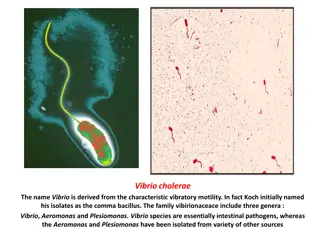
SWINE FEVER (Hog Cholera) Synonym:- Hog cholera ,Swine plague, Classical swine fever, European swine fever. The disease causes multimillion dollar losses before eradication programme has been taken. USA, UK, Australia, New Zealand, Ireland eradicated these disease. But again the disease came in some of the countries, like Belgium, Netherland, Germany etc in 1990.
SWINE FEVER (Hog Cholera) Swine fever is an acute contagious viral disease of pigs caused by pestivirus and characterized by gluing of eyes, button ulcers in intestine, congestion and haemorrhage in visceral organs. 2
Etiology The virus of Hog cholera and Mucosal disease are antigenically similar. Pestivirus of Togaviridae family RNA virus
Incubation period 1 to 4 days by artificial infection and 7 days in natural infection
Incidence First recognized in 1885 in the United States, its viral aetiology was established in 1903 The disease is seen worldwide including India Susceptibility The pig is the only domestic animal which is naturally infected by the virus
Transmission The infection is usually acquired by ingestion, but inhalation is also a possible route Routes of infection Digestive tract Respiratory tract Conjunctiva Nasal mucosa
Pathogenesis The first virus enters into blood after multiplication in tonsillar tissue After that the virus will infect the lymphoid organs and also infect liver, spleen and other internal organs. There is leukopaenia, thrombocytosis.
Sing & Symptoms The virus has special affinity for the endothelial cells of blood vessels & reticulo -endothelial system. The vascular endothelial cells swell, proliferate and occlude the lumen. The wall of blood vessels undergo hyaline degeneration with infiltration by lymphocytes, macrophages and plasma cells. These changes of blood vessels are the cause of hemorrhages, necrosis and infarection found in various organs.
Spleen severally infarcts are wedge shaped, found on the edges of the orgen & are browinesh in color. Lungs croupous pneumonia. In the Intestine especially in the cecum and colon, are found the characteristic Button shaped ulcer . Kidney found petechiae on the cortex extending deeply into the parenchyma. These gives a characteristic Turkey egg appearance .
Skin shows erythematous patches which become cyanotic. Later vesicles may form on the lips, vulva & edges of ears. Brain non purulent meningo encephalomyelitis. Perivascular cuffing - i.e accumulation of lymphocytes, monocyes, plasma cells & local histocytes in the perivascular space ( Robin Virchow).
Sing & Symptoms In mixed infections Salmonella cholerae suis often complicates the picture. The original name of the disease Hog Cholera was derived from the discovery.
Clinical Signs Acute disease Huddling, dullness High fever (105oF) Anorexia Erythema, cyanosis Petechiae Staggering, weakness Convulsions Poor reproductive performance Abortions, stillbirths Deformities
Post Mortem Lesions Highly variable Acute infection Hemorrhage Necrotic foci in tonsils Petechiae Kidney, larynx, trachea, intestines, spleen, lungs
Post-Mortem Lesions Chronic infection Necrotic foci Intestinal mucosa ( button ulcers) Epiglottis Larynx Congenital infection Cerebellar hypoplasia, thymic atrophy, deformities of head and legs Photo courtesy of Dr. R. Panciera, Oklahoma State University
Differential Diagnosis Diagnosis is impossible without lab testing Porcine reproductive and respiratory syndrome (PRRS) Porcine circovirus associated disease Salmonellosis Erysipelas Leptospirosis Aujeszky s disease (pseudorabies) African swine fever Tonsil samples should be sent with every submission to your state diagnostic lab
Sampling If Classical Swine Fever is suspected: The proper animal health authorities should be contacted, before collecting or sending any samples, Samples should only be sent under secure conditions, to authorized laboratories to prevent spread of the disease
Diagnosis Laboratory Tests Detect virus, antigens, nucleic acids Tissue samples (tonsils, spleen, kidneys, distal ileum) Whole blood ELISA or direct immunofluorescence Serology ELISA or virus neutralization Comparative neutralization test Definitive test
Disease Control Disinfectants Sodium hypochlorite Phenolic compounds Virus sensitive to Drying Ultraviolet light pH of less than 3 or greater than 11 Killed at high temperatures 150oF for 30 minutes; 160oF 1 minute
")
")