Syringomyelia, Brown-Séquard Syndrome, and Motor Neuron Disease
Learn about Syringomyelia, Brown-Séquard Syndrome, and Motor Neuron Disease - their symptoms, causes, and clinical features. Explore images and detailed descriptions of these spinal cord conditions, including their implications and management.

Download Presentation

Please find below an Image/Link to download the presentation.
The content on the website is provided AS IS for your information and personal use only. It may not be sold, licensed, or shared on other websites without obtaining consent from the author. If you encounter any issues during the download, it is possible that the publisher has removed the file from their server.
You are allowed to download the files provided on this website for personal or commercial use, subject to the condition that they are used lawfully. All files are the property of their respective owners.
The content on the website is provided AS IS for your information and personal use only. It may not be sold, licensed, or shared on other websites without obtaining consent from the author.
E N D
Presentation Transcript
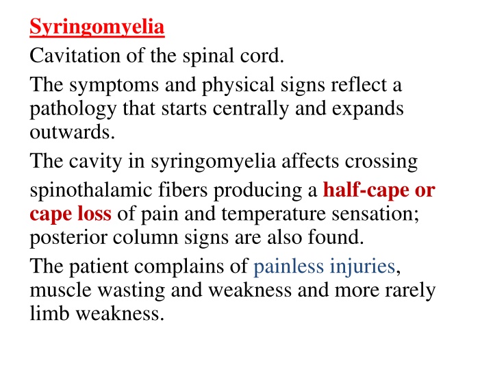
Syringomyelia Cavitation of the spinal cord. The symptoms and physical signs reflect a pathology that starts centrally and expands outwards. The cavity in syringomyelia affects crossing spinothalamic fibers producing a half-cape or cape loss of pain and temperature sensation; posterior column signs are also found. The patient complains of painless injuries, muscle wasting and weakness and more rarely limb weakness.
Generally, there are two forms of syringomyelia: congenital Arnold Chiari malformation acquired the second major form of syringomyelia occurs as a complication of trauma, meningitis, hemorrhage, tumor.
Clinical feature There is amyotrophy(LMN) at the level of the cavity with tendon reflex loss. In advanced stages Charcot joints develop. Below the cavity there may be upper motor neuron symptoms and signs and disturbances of sphincter function, which contrast with the lower motor neuron symptoms and signs at the level of the syrinx.
Brown- Sequard Syndrome Brown- S quard syndrome usually follows spinal cord hemisection as a result of penetrating trauma can also occur with large disc herniation spinal epidural hematoma. The classic description involves a dissociated sensory loss with contralateral loss of pain and temperature but preserved ipsilateral light touch and posterior column function. In addition, there is ipsilateral motor paralysis below the level of the lesion.
(MND) Motor neuron disease Motor neuron disease e.g.(amyotrophic lateral sclerosis) is a progressive neuronal degenerative disease that leads to severe disability and death begins usually above the ages of 50years . It is characterized by degeneration of anterior horn cells in the spinal cord, motor nuclei of the lower cranial nerves in the brainstem, and corticospinal and corticobulbar pathways.
it is a disease in which certain nerve cells in the brain and spinal cord slowly die. These nerve cells are called motor neurons, and they control the muscles that allow you to move the parts of your body. People who have MND gradually become more disabled, how quickly the disease gets worse is different for everyone. Some people live with ALS for several years. But over time, ALS makes it hard to walk, speak, eat, swallow, and breathe.
So there is features of combination of upper motor and lower motor type (characterized clinically by wasting, weakness and fasciculation of the affected muscles with hyperreflexia.
PATTERNS OF INVOLVEMENT OF MOTOR NEURON DISEASE Progressive muscular atrophy Predominantly spinal motor neurons affected Weakness and wasting of distal limb muscles at first Fasciculation in muscles Tendon reflexes may be absent Progressive bulbar palsy Early involvement of tongue, palate and pharyngeal muscles Dysarthria/dysphagia Wasting and fasciculation of tongue May be pyramidal signs as well
Amyotrophic lateral sclerosis Combination of distal and proximal muscle-wasting and weakness, fasciculation . Spasticity, exaggerated reflexes, extensor plantars Bulbar and pseudobulbar palsy follow eventually Pyramidal tract features may predominate NOTE NO SENSORY IVOLVEMENT NO SPHINCTER DYSFUNCTION NO OCULAR MUSCLES AFFECTION NO CEREBELLAR INVOLVEMENT
INVESTIGATION the clinical features are highly suggestive Electromyography helps to confirm the presence of fasciculation and denervation. Spinal imaging and brain scanning may be necessary to exclude focal spinal or cerebral disease. CSF examination is usually normal.
Management riluzole, has recently been shown to have a small effect in prolonging life expectancy by about two months . Psychological and physical support. Prognosis the mean time from diagnosis to death is 1 year, with most patients dying within 3-5 years of the onset of symptoms


 Motor neuron disease")