The Heart's Electrical System: Understanding Cardiac Conduction
The heart's electrical system controls its rhythm and coordination. From the Sinoatrial node to the Purkinje fibers, each component plays a crucial role in ensuring proper heart function. Explore how electrical impulses are generated and conducted through the heart, allowing for efficient blood circulation and coordination between atria and ventricles.

Download Presentation

Please find below an Image/Link to download the presentation.
The content on the website is provided AS IS for your information and personal use only. It may not be sold, licensed, or shared on other websites without obtaining consent from the author.If you encounter any issues during the download, it is possible that the publisher has removed the file from their server.
You are allowed to download the files provided on this website for personal or commercial use, subject to the condition that they are used lawfully. All files are the property of their respective owners.
The content on the website is provided AS IS for your information and personal use only. It may not be sold, licensed, or shared on other websites without obtaining consent from the author.
E N D
Presentation Transcript
Prof. Mona Soliman, MBBS, MSc, PhD Head, Medical Education Department Professor of Physiology and Medical Education Chair of Cardiovascular Block College of Medicine King Saud University
Cardiac Electrical Activity Automaticity of the heart: the heart is capable of 1. Generating rhythmical electrical impulses 2. Conduct the impulses rapidly through the heart The atria contract about one sixth of a second ahead of ventricular contraction Why? To allows filling of the ventricles before they pump the blood into the circulation
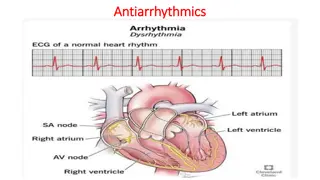
The Specialized Excitatory and Conductive System of the Heart The sinoatrial (S-A node) The internodal pathway The atrioventricular (A-V node) The atrioventricular bundle (Bundle of His) Purkinje fibers 1. 2. 3. 4. 5.
Conduction of Impulses Sinoatrial node (S-A node): Located in the superior lateral wall of the right atrium near the opening of the superior vena cava Pacemaker of the heart Why? Its rate of rhythmic discharge is greater than any other part in the heart Highest frequency Is capable of originating action potentials
Conduction of Impulses Atrioventricular (A-V) node: Located in the posterior wall of the right atrium Delay in the conduction of impulses (0.1 sec) Why? Allows time for the atria to empty the blood into the ventricles before ventricular contraction begin
Conduction of Impulses The Purkinje System Purkinje fibers are very large fibers Transmit action potentials at a very high velocity (0.1-4.0 m/sec) Why? very high permeability of gap junctions ions are transmitted easily from one cell to the next enhance the velocity of transmission Ventricular muscle contract at almost the same time
Conduction of Impulses The Purkinje System Penetrate atrioventricular fibrous tissue divides into right and left bundle branches each branch spread toward the apex of the heart divide into small branches penetrate and become continuous with cardiac muscle fibers
Control of Excitation and Conduction in the Heart The impulse normally arise s in the sinus node Why? The Sinus Node is the Pacemaker of the Heart Its rate of rhythmical discharge is faster than that of any other part of the heart
Abnormal Pacemakers Ectopic pacemaker: a pacemaker elsewhere than the sinus node The cause: Any other part of the heart develops a rhythmical discharge rate that is more rapid than that of the sinus node Example: the A-V node or in the Purkinje fibers 1.
Abnormal Pacemakers 2. Blockage of transmission of the cardiac impulse from the sinus node to the other parts of the heart Example: A-V block cardiac impulses fails to pass from atria into the ventricles the atria continues to beat at the normal rate of rhythm of the S-A node a new pacemaker develops in the Purkinje system with a new rate
Control of Heart Rhythmicity and Impulse Conduction by the Cardiac Nerves The heart is supplied with both sympathetic and parasympathetic nerves Parasympathetic nerves (vagi): mainly to the S-A and A-V nodes Sympathetic nerves: all parts of the heart with strong supply to the ventricles
Parasympathetic stimulation of the heart rate of rhythm of the S-A node transmission of impulses to the A-V node Strong stimulation of the vagi: Stop completely the rhythmical excitation by the S-A node Block completely transmission of cardiac impulses from the atria to the ventricle Some point in the Purkinje fibers develops a rhythm of its own Ventricular Escape
Sympathetic stimulation of the heart rate of rhythm of the S-A node transmission of impulses to the A-V node force of contraction
For further readings and diagrams: Textbook of Medical Physiology by Guyton & Hall Chapter 10 (Rhythmical Excitation of the Heart)