The Human Eye: Understanding Glaucoma and Eye Anatomy
The human eye comprises intricate structures like the fibrous coat, vascular pigmented coat, and nervous coat. Glaucoma, a condition affecting the optic nerve, can lead to vision loss if not managed properly. Explore the layers of the eye and understand how conditions like glaucoma impact eye health.

Download Presentation

Please find below an Image/Link to download the presentation.
The content on the website is provided AS IS for your information and personal use only. It may not be sold, licensed, or shared on other websites without obtaining consent from the author.If you encounter any issues during the download, it is possible that the publisher has removed the file from their server.
You are allowed to download the files provided on this website for personal or commercial use, subject to the condition that they are used lawfully. All files are the property of their respective owners.
The content on the website is provided AS IS for your information and personal use only. It may not be sold, licensed, or shared on other websites without obtaining consent from the author.
E N D
Presentation Transcript
DRUGS AND HUMAN EYE: GLAUCOMA BY SHAEMA M. ALI
The eye Coats of the eyeball The eyeball consist of three coats 1- the fibrous coat 2- the vascular pigmented coat 3- the nervous coat
Fibrous coat The fibrous coat is made up of a posterior opaque part, the sclera, and an anterior transparent part, the cornea.
Posteriorly, the sclera is pierced by the optic nerve and is fused with the dural sheath of that nerve in an area called lamina cribrosa at the optic disc. It is relatively week area and can be made to bulge into the eyeball by a rise of cerebrospinal fluid pressure in the subarachinoid space
If the IOP rises, the lamina cribrosa will bulge outward, producing a cupped disc as seen through the opthalmoscope.
Vascular pigmented coat The vascular pigmented coat consists from behind forward of the coroid, the ciliary body and the iris.
The choroid is composed of an outer pigmented layer and an inner highly vascular layer.
The ciliary body composed of ciliary processes ( secretion of aqueous humor) and ciliary muscles ( contraction of these muscles helps in accomodation for near vision).
The iris is a thin contractile pigmented diaphram with a central aperture, the pupil It is suspended in the aqueous humor between the cornea and the lens It divides the space between the cornea and the lens into an anterior and a posterior chamber
The muscle fibers of the iris are involuntary and consist of circular( parasympathetic innervation; miosis) and radiating fibers (sympathetic innervation; mydriasis).
Nervous coat: the retina The retina consist of an outer pigmented layer and an inner nerveus layer
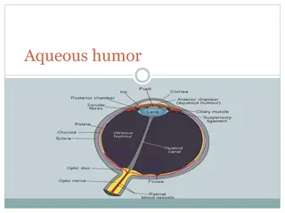
Contents of the eyeball -lens biconvex body situated behind the iris and in front of the vitreous body It attached to the ciliary processes by the suspensory ligament. -Fluid system of the eye- Introcular fluid The eye is filled with intraocular fluid which maintain sufficient pressure in the eyeball to keep it distended.
This fluid can be divided into two portions Aqueous humor, which lies in front of the lens and vitreous humor , which is between the posterior surface of the lens and the retina.
Formation and outflow of aqueous humor The aqueous humor is a clear fluid that fills the anterior and posterior chambers of the eyeball It is a secretion or transudate from the ciliary processes, from which it enters the posterior chamber.
It then flows into the anterior chamber through the pupil and is drained away through the spaces at the iridocorneal angle into the canal of schlemm.
Obstruction to the draining of the aqueous humor results in a rise in IOP, called glucoma. This can produce degenerative changes in the retina, with consequent blindness.
The disease is usually classified according to the manner in which aqueous humor outflow is impaired. - Primary open-angle glucoma - Primary angle-closure glucoma
Primary open-angle glucoma (POAG) POAG is associated with a relative obstruction to aqueous outflow through the trabecular meshwork and is a chronic progressive disease of insidious onset usually affecting both eyes.
Primary angle closure glucoma (PACG) PACG is a condtition in which closure of the angle by the peripheral iris results in a reduction in aqueous outflow. It occurs in predisposed eyes and is frequently unilateral.
Etiology The factors that determine the level of IOP are the rate of aqueous humor production and the resistance encountered in the outflow channels. A fine balance between these is necessary to keep the pressure within the eye within the range of 16-21 mmHg.
Clinical manifestation POAG, because of its insidious onset, is usually asymptomatic until it has caused a significant loss of visual field. The earliest clinically significant field defect is scotoma, which is an area of depressed vision within the visual field.
Acute PACG is due to sudden closure of the angle and a severe elevation in IOP. The symptoms include rapidly progressive visual impairment, periocular pain and congestion of the eye. In severe cases nausea and vomiting may occur
Investigations IOP is measured by tonometry Scotoma can be detected by using perimetry Gonioscopy is used to estimate the width of the chamber angle. Cupping of the optic disc seen by ophthalmoscopy is used in both diagnosis and assessment of the efficacy of treatment.
Treatment Primary angle glucoma The prostaglandin analougues latanoprost and travoprost and the prostamide bimatoprost are indicated for first line use, as are the B blockers as these produce the greatest fall in IOP.
Carbonic anhydrase inhibitors and sympathomimetics which results in smaller fall in IOP, are considered second line drugs.
Pilocarpine is usually reserved for those patients not controlled by a combination of the drugs listed above. Oral therapy with carbonic anhydrase inhibtors is usually reserved for use as the final stage of treatment in those complex glaucomas awaiting surgery.
Primary angle closure glaucoma Analgesics and antiemitcs are sometimes needed. Intensive miotic therapy is usually ineffective and the IOP needs to be lowered by drugs that reduce aqueous humor production.
An intravenous loading dose of acetazolamide followed by oral treatment should allow the IOP to drop sufficiently to relieve iris ischemia and allow the sphincter to respond to pilocarpine therapy.
If this fails to decrease IOP, hyperosmotic agents ( glycerol and mannitol) may be required. Once the IOP has been reduced medically, the condition is usually treated surgically.
Drug induced glaucoma Several different drugs have the potential to cause the elevation of IOP which can occur via an open angle mechanism or a closed angle mechanism.
Steroid-induced glaucoma is a form of open angle glaucoma that usually associated with topical steroid use.
medications that have a direct or secondary effect, either to stimulate sympathetic or inhibit parasympathetic activation causing pupillary dilation can precipitate acute ACG in patients with occludable angles.
the most frequently performed surgical procedures create a fistula to act as a new route for aqueous outflow. Laser trabeculoplasty Laser ciliary ablation
These include topical anticholinergic or sympathomimetic dilating drops, tricyclic antidepressant, monoamine oxidase inhibitors, antihistamines, antiparkinsonian drugs, antipsychotic medication and antispasmolytic agents.
Note: sulfa containig medications may induce ACG by a different angle closure mechanism involving anterior rotation of the ciliary body.


")
")
")