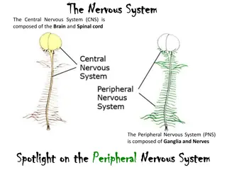
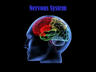
The Nervous System: Organization and Function
Explore the intricate workings of the nervous system, including its general organization, supporting cells like astrocytes and microglia, and the structure of neurons. Learn about the important roles of neuroglia cells, myelin sheath, and different types of neurons in transmitting and processing information throughout the body.

Download Presentation

Please find below an Image/Link to download the presentation.
The content on the website is provided AS IS for your information and personal use only. It may not be sold, licensed, or shared on other websites without obtaining consent from the author. If you encounter any issues during the download, it is possible that the publisher has removed the file from their server.
You are allowed to download the files provided on this website for personal or commercial use, subject to the condition that they are used lawfully. All files are the property of their respective owners.
The content on the website is provided AS IS for your information and personal use only. It may not be sold, licensed, or shared on other websites without obtaining consent from the author.
E N D
Presentation Transcript
I. General organization of nervous system A. CNS 1. brain 2. spinal cord B. PNS 1. sensory 2. motor a. Somatic b. ANS -sympathetic -parasympathetic
II. Nervous Supporting Cells - neuroglia A. Astrocytes 1. Connect to capillaries 2. Mopping up chemical environment of brain as far as potassium ions and neurotransmitters 3. Help to create blood brain barrier
B. Microglia 1. spider-like phagocytes 2. debris, dead brain cells, bacteria
C. Ependymal cells 1. lines cavities in CNS 2. beating of cilia moves cerebrospinal fluid 3. fluid nourishes and cushions CNS 4. creates CSF in the choroid plexi of the brain s ventricles
D. Oligodendrocytes 1. wrap axons of several nerve cells with fatty layer 2. produces myelin sheath 3. speeds conduction 4. located with the CNS
E. Schwann cells 1. located outside of CNS 2. produce myelin sheath as do the oligodendrocytes 3. takes several Schwann cells to produce the myelin sheath for one axon of one nerve cell
F. Glia cells in general 1. resemble neurons 2. not excitable 3. supportive cells 4. capable of repeated mitosis 5. gliomas-glial tumors
III. Neurons A. Structure 1. cell body 2. nissl bodies-rer 3. dendrites 4. axon 5. axon hillock 6. axon collateral 7. axon terminals 8. neurotransmitters 9. synaptic cleft
B. Myelin sheath 1. functions 2. PNS-Schwann cell 3. Node of Ranvier 4. Can form a pathway for regrowth of damaged axon 5. multiple sclerosis
C. Neurons classified by function 1. afferent 2. interneuron 3. efferent 4. ganglia 5. nuclei 6. gray matter 7. white matter
D. Neurons classified by structure 1. multipolar- most common 2. bipolar- located in some sensory organs such as the eye 3. unipolar- sensory neuron
IV. Neuron physiology A. Membrane traits 1. semipermeable 2. Na/K ion pump 3. Leak gates 4. gated channels a. Ligand-gated b. Voltage-gated Membrane of a certain cell
B. Resting membrane characteristics 1. semipermeable 2. negative charged proteins 3. relatively impermeable to Na and Cl ions 4. bit more permeable to K ions 5. due to action of Na/K ion pump notice separation of ions 6. potassium ions leak out due to K ion leak channels
C. Resting membrane potential 1. at rest, interior of cell possesses slightly negative charge 2. -70 mV 3. due to K ion movement mainly 4. diffusion out 5. electrical attraction in 6. slightly more positive charge outside 7.http://www.youtube. com/watch?v=YP_P6b YvEjE
D. Changing the resting membrane potential in a resting neuron 1. depolarization 2. hyperpolarization 3. changes in extracellular K ions (hypokalemia) 4. changes in extracellular Na ions 5. changes in extracellular Ca ions a. Ca ions are attracted to negative proteins of Na gated channels b. If Ca ion concentration falls-fewer Ca ions attached to Na gated channels-causes channels to open- produces???hypocalcemia c. If Ca ion levels rise-???
E. Graded potentials 1. strictly local event 2. caused by change in local ion gates 3. change brought about by several possible stimulus sources 4. chemical, voltage changes, temperature, mechanical stimulation 5. may be excitatory or inhibitory 6. conducted but in a decremental manner
F. Action potential 1. produced by graded potentials 2. threshold potential 3. intiates series of membrane gate changes 4. wave of depolarization 5. repolarization 6. hyperpolarization 7. return to normal 8. all-or-none 9. https://highered.mcgraw- hill.com/sites/007249585 5/student_view0/chapter 14/animation__the_nerve _impulse.html
G. Refractory period 1. definition 2. absolute 3. relative
H. Frequency carries information 1. action potentials don t vary in magnitude 2. threshold stimulus produces one action potential 3. submaximal stimuli produce increasing frequency of action potentials until 4. maximal stimulus-lowest stimulus strength that produces maximum frequency of action potentials 5. supramaximal stimulus
I. Propagation of action potentials 1. concentration difference of ions on either side of membrane represents potential energy-kind of like of cocked gun 2. stacked dominoes waiting to fall over 3. one domino falling over initiates a wave of action potentials spreading out like the ripples in a pond 4. each action potential is just as strong as the previous action potential 5. strength does not diminish as nerve impulse moves down the axon 6. http://highered.mcgraw- hill.com/sites/9834092339/student_view0/chapter44/action_poten tial_propagation_in_an_unmyelinated_axon.html 7. http://www.youtube.com/watch?v=DJe3_3XsBOg
V. Synapses A. Anatomy 1. presynaptic membrane 2. synaptic cleft 3. postsynaptic membrane 4. synaptic vesicles 5. receptor sites for transmitter substance
B. Physiology of synapse 1. action potential arrives 2. Calcium ion channels open 3. synaptic vesicles fuse with membrane 4. transmitter substance released 5. diffusion of transmitter substance 6. binding to receptors 7. creates a graded potential 8. may bring postsynaptic membrane to threshold 9. nerve gas-blocks cholinesterase 10. IPSP or EPSP
C. You tube of synaptic events http://www.youtube.com/watch?v=LT3VKAr4r oo
D. Types of Synapses 1. axo-dendritic 2. axo-somatic 3. axo-axonic a. Presynaptic inhibition of enkephalins and endorphins in brain sensory neurons blocking Ca channels b. Presynaptic facilitation due to serotonin release- causes Ca channels to open
E. Post synaptic fiber as a neural integrator 1. temporal summation 2. spatial summation 3. neural integrator