The Physiology of Proprioception in Balance
Identify sensory receptors, pathways, roles of dorsal column system, and spinocerebellar tract in proprioception. Learn about sensory vs. motor ataxia, nervous system organization, dermatomes, and types of proprioceptors. Discover how proprioception informs body position, movement, muscle tension, and more.

Download Presentation

Please find below an Image/Link to download the presentation.
The content on the website is provided AS IS for your information and personal use only. It may not be sold, licensed, or shared on other websites without obtaining consent from the author.If you encounter any issues during the download, it is possible that the publisher has removed the file from their server.
You are allowed to download the files provided on this website for personal or commercial use, subject to the condition that they are used lawfully. All files are the property of their respective owners.
The content on the website is provided AS IS for your information and personal use only. It may not be sold, licensed, or shared on other websites without obtaining consent from the author.
E N D
Presentation Transcript
Text Important Formulas Numbers Doctor notes Notes and explanation Lecture No.14 Only Those Who Dare To Fail Greatly Can Ever Achieve Greatly 1
Physiology of Proprioception in Physiology of Proprioception in Balance Balance Objectives: 1. 2. 3. Identify the major sensory receptors & pathways. Describe the components, processes and functions of the sensory pathways. Appreciate the dorsal column system in consciousproprioception (anatomy and functions). Describe the spinocerebellar tract pathway in unconscious proprioception from muscles, tendons, and joints. Defferentiate between sensory and motor ataxia. 4. 5. 2
ONLY IN FEMALES SLIDES Organization of the Nervous System (introduction) The brain Central Nervous System (the center of integration and control) The spinal cord Two big initial divisions: 31 Spinal nerves: carry info to and from the spinal cord. Peripheral Nervous System (the nervous system outside of the brain and spinal cord) 12 Cranial nerves: carry info to and from the brain. 3
Dermatomes Dermatome is an area on skin supplied by a single spinal nerve. 4
What is Proprioception ? ONLY IN MALES SLIDES ONLY IN FEMALES SLIDES Proprioception stems from the latin word proprius which Encapsulated Nerve Endings. Monitor stretch in locomotory organs. means "one's own or "individual . It is the sense of one`s own body position. It is also called proprioceptive / position sense. It is the awareness of body position and of movements of body parts. Strength of effort being employed in movements. ONLY IN MALES SLIDES types of proprioceptors: Exteroception: By which one perceives the outside world. 1. Interoception: By which one perceives pain, hunger, and the movement of internal organs. 2. It can be divided into: Static proprioception: conscious perception of the orientation of the different parts of the body with respect to one another. 1. Dynamic proprioception: rate of movement sense (also called kinesthesia). 2. 5
Exrta Cont. Proprioception informs us about: The location of a body part in relation to other parts. The rate of movement of a body part when it is moving. The degree to which our muscle are being contracted or stretched. The amount of tension created in our tendons. The head orientation in relation to the ground and in response to movement. Proprioceptive information is carried from periphery to the CNS by proprioceptors and other somatic receptors. 6
Types of Proprioceptors This slide is important Muscle spindles also detect the degree of strength on the muscles and rate of change of movement. Golgi tendon is stimulated when there is overstretch : a stretch that may tear the muscle or separate it from it's tendon. Proprioceptors include the muscle spindles, Golgi tendon organs, and joint receptors. These provide a sense of body position and allow fine control of skeletal movements. Muscle spindles: 1. Detect how much a muscle is stretched. Imbedded in the perimysium between muscle fascicles. Golgi tendon organs: 2. Located near the muscle-tendon junction. Detect tension of a muscle on its tendon. Provide information about the strength of contraction & tension. Joint Kinesthetic receptors: 3. Are mechanoreceptors + sensory (nerve endings) in the joint capsules, they detect angle and movement of the joints. N.B: Cutaneous & deep receptors also contribute to proprioception 7
ONLY IN MALES SLIDES Cont. There are two types: These are main ascending sensory pathways for proprioception. Types of Proprioception Conscious proprioception Unconscious proprioception is communicated to the cerebellum primarily via: It reaches the level of sensory cerebral cortex (cerebrum) via the dorsal column tract. The dorsal spino-cerebellar tract (dSCT). 1. The ventral spino-cerebellar tract (vSCT). 2. 8
ONLY IN MALES SLIDES Sensory receptors types Mechanoreceptors: which detect mechanical compression or stretching of the receptor or of tissues adjacent to the receptor, ex: proprioceptors. Thermoreceptors: which detect changes in temperature, some receptors detecting cold and others warmth. Nociceptors (pain receptors): which detect damage occurring in the tissues, whether physical damage or chemical damage, ex: free nerve endings. Electromagnetic receptors: which detect light on the retina of the eye, ex: rods and cones. Chemoreceptors: which detect taste in the mouth, smell in the nose, oxygen level in the arterial blood, osmolality of the body fluids, carbon dioxide concentration, and perhaps other factors that make up the chemistry of the body, ex: chemo R in carotid bodies 9
Classification of sensory receptors ALL this smart art is extra Classification of Sensory Receptors A-Based on their location (Sherrington 1906) B-Based on their adequate stimulus concerned with the external environment -Found on the the surface of the body -Monitor changes in the external environment Exteroceptors Thermoreceptors (cold & heat) Photoreceptors (in the retina). Nociceptors (pain receptors) Chemoreceptors (ex: taste receptors) Mechanoreceptors concerned with the internal environment (visceral) Interoceptors concerned with one`s own body position -Are those relating to the physical state of the body, including joint position, and sate of tendons and muscles. Sensory receptors classified according to: Location Type of stimulus detected Structure ONLY IN FEMALES SLIDES Proprioceptors 10
Cont. Extra ONLY IN MALES SLIDES Adaptation of receptors: Based on their speed of adaptation When a continuous sensory stimulus is applied, the receptor responds at a high impulse rate at first and then at a progressively slower rate until finally the rate of action potentials decreases to very few or often to none at all. Rabidly adapting (RA) Slowly adapting (SA) Fire upon stimulus and cease firing when the stimulus remains constant (phasic receptors). Getting used to the bad smell in a room. Being aware of a ring or a watch only when putting them on or taking them off (onset and offset). Fire as long as the stimulus is there (tonic receptors). Nociceptors and proprioceptors show no or very little adaptation. 11
ONLY IN MALES SLIDES Cont. Adaptation of receptors Receptor potential of the pacinian corpuscle: Rapid adapting or phasicreceptors. Ex: meissner s corpuscles(touch), pacinian corpuscles(vibration) Slowly adapting or tonic receptors. Ex: ruffini s (pressure ,skin stretch ) krause s end bulbs, and merkel s disks. Non adapting receptors. Ex: free nerve endings for pain sensation. For joint position and vibration sensation (Also Ruffini s endings). The receptor potential produced by compression induces a local circuit of current flow that spreads along nerve fiber. The frequency of repetitive action potentials transmitted from sensory receptors increases approximately in proportion to the increase in receptor potential. Mechanisms by which receptors adapt: First, the pacinian corpuscle is a viscoelastic structure so that after stimulation within few hundredths of a second, the fluid within the corpuscle redistributes, so that the receptor potential is no longer elicited. The second mechanism of adaptation of the pacinian corpuscle, but a much slower one, results from accommodation , which occurs in the nerve fiber itself. This probably results from progressive inactivation of the sodium channels in the nerve fiber membrane. The Explanation of this picture next slide. 12
Doctors explanation when deformed area is compressed it cause a receptor potential if this receptor potential is enough it causes action potential. receptor potential is localized graded and doesn't spread. AP: it's either yes or no ( not graded ) and spreads. If you want to stimulate them in a continuous state you increase the strength of stimulus. Pain receptors don't adapt and proprioceptors also. 13
ONLY IN MALES SLIDES Structure of proprioception Structures concerned with proprioception: Proprioceptors (spatial orientation, four inputs). Brain Stem (Cortico, Rubro, Vestibulo, Reticulo, Olivo, Tectospinal). vestibular system (apparatus, nuclei). Ascending Tracts. Visual system. Cerebellum (flocculonodular lobe dynamic equilibrium, Uvula Static equilibrium). Conscious perception will be perceived by cortex so impulses passes through brain stem. Reticulo: you must be awake to perceive the perception. Olivo: because it's connected to cerebellum. When all equilibrium contributers are damaged the person still can stand and maintain balance because of visual clues if this person closes his eyes he falls. Uvula responsible for static equilibrium and flocculonodular for dynamic. Cerebral cortex (primary cortical center for equilibrium located in the parietal lobe deep in the sylvian fissure). 14
The doctor was explaining this Muscle Spindles & Golgi Tendon Organs Muscle spindles: They detect changes in the length of 1. muscle. They convey length information to the CNS via group I and II afferent neurons. This information is important for determining the position of body part. Golgi tendon organs They detect changes in muscle tension. 2. 15
ONLY IN FEMALES SLIDES Classification of nerve fibers An Overview of Sensory Pathways and the Somatic Nervous System. Neural pathways: Afferent pathways: Sensory information coming from the sensory receptors through peripheral nerves to the spinal cord and to the brain . Efferent pathways: Motor commands coming from the brain and spinal cord, through peripheral nerves to effecter organs . 16
Unencapsulated nerve endings Encapsulated Encapsulated Nerve Endings: Consist of one or more end fibers of sensory neurons. Enclosed in connective tissue. Include four main types: Meissner s corpuscles 1. Pacinian corpuscles 2. Ruffini s corpuscles 3. Proprioceptors 4. 17
Cont. Encapsulated nerve endings This slide is important 18
ONLY IN MALES SLIDES Sensory modalities 19
ONLY IN MALES SLIDES Reflex arc Stimulus Afferent Center Efferent Effector Effect Receptor Inverse stretch reflex Stretch reflex Muscle stretch Muscle spindle Golgi organ tendon Afferent 1a Over stretch Afferent 1b Efferent motor Efferent Motor Extrafusal fibers Spinal cord (center) Inhibitory interneuron Spinal cord (center) Reciprocal inhibition of antagonist Reciprocal inhibition of antagonist Muscle contraction Extrafusal fibers Muscle relaxation 20
ONLY IN MALES SLIDES Types of mammalian fibres 21
ONLY IN MALES SLIDES Classification of sensory nerve fibres 22
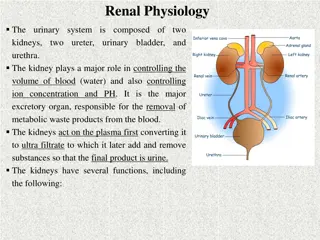
Spinal cord tracts Sensory information from receptors throughout most of the body is relayed to the brain by means of ascending tracts of fibers that conduct impulses up the spinal cord. When the brain directs motor activities, these directions are in the form of nerve impulses that travel down the spinal cord in descending tracts of fibers. Spinal cord tracts Ascending tracts (sensory) Descending tracts (motor) A Cross-section view of spinal cord- wider laterllay than anteroposteriorly. In the middle on the dorsal side is a shallow groove called the posterior median sulcus and on the ventral side is the anterior median fissure (deeper). center consist of gray matter shaped like a butterfly and there is an opening at the center. Spinal cord is protected by three layers of meninges. The only difference from the brain is that the dural matter does not attach to bone. The dural matter is surrounded externally by a layer of cushioning fat called epidural space. ONLY IN FEMALES SLIDES 23
Organization of spinal cord Grey matter: organization White matter in the spinal cord Fibers run in three directions, ascending, descending and transversely. Dorsal half sensory roots and ganglia. Ventral half motor roots. Divided into three funiculi (columns), posterior, lateral and anterior. Dorsal and ventral roots fuse laterally to form spinal nerves. Each funiculus contains several fiber tracks. Four zones are evident within the gray matter somatic sensory (SS), visceral sensory (VS), visceral motor (VM), and somatic motor (SM). Fiber tract names reveal their origin and destination. Fiber tracts are composed of axons with similar functions. 24
Ascending sensory tracts Sensory pathways: 3 neurons: 1st: enters spinal cord from periphery. 2nd: crosses over (decussates), ascends in spinal cord to thalamus. 3rd: projects to somatosensory cortex. Spinal tracts: These are known as sensory and motor pathways consisting of multineuron pathways connecting the CNS to the PNS. At some point most pathways crossover (decussate), Ascending (sensory) Pathways : Dorsal column pathway: carries signal of fine touch, pressure, vibration , stereognosis and concious proprioception, ascends up dorsal white column in fasciculus gracilis or cutaneatus to medulla oblongata to the thalamus to primary somatosensory cortex (post central gyrus). Posterior and anterior spinocerebellar pathways: carry subsconcious proprioception. Dorsal gray horn to lateral column to medulla oblongata to pons to cerebellum. Spinothalamic pathway: carries signals of pain, temperature, deep pressure, and course touch. From psterior gray horn decussate into lateral and anterior funiculi up to the thalamus to primary somatosensory cortex (postcentral gyrus). There are several ascending sensory systems. Dorsal column system & Anterolateral system. Each system carries different types of sensations or modalities proprioception, pain, temperature, fine touch, crude touch, vibration. Sensory systems allow us to detect, analyze and respond to our environment . Ascending pathways carry information from sensory receptors to the brain. 1. Conscious: reach cerebral cortex. Unconscious: do not reach cerebral cortex. Sensations from body reach the opposite side of the brain. 2. Stereognsis the mental perception of depth or three dimensionality by the senses, usually in reference to the ability to perceive the form of solid objects by touch. 3. Conscious feeling always relayed to cerebral cortex so if only cerebellar tracts are intact you won't feel any thing. 25
ONLY IN MALES SLIDES Cont. Anterolateral system Ventral and lateral Spinothalamic lesion spinothalamic tracts. Pain. Thermal sensations, (warmth & cold). Crude touch and pressure sensations capable only of crude localizing ability on the surface of the body. Tickle and itch sensations. Sexual sensations. 26
Dorsal columnmedial lemniscal system Touch sensations requiring a high degree of localization of the stimulus. Touch sensations requiring transmission of fine gradations of intensity. Carries fine touch, tow point discrimination, pressure, vibration and conscious proprioception signals. Phasic sensations like vibratory sensations. Sensations that signal movement against skin. Joints Position sensations (Proprioception). Pressure sensations requiring fine degrees of judgment of intensity. Strereognosis. FineTouch Pressure Vibration Position Movement 3rd neuron Projects to somatosensory cortex. Fact 2nd neuron Crosses over in medulla, ascends to thalamus. conducting A / -fiber neurons 1st neuron Enters spinal cord through dorsal root, ascends to medulla (brain stem). 27
ONLY IN MALES SLIDES Proprioception from head In this pathway through the brain stem, each medial lemniscus is joined by additional fibers from the sensory nuclei of the trigeminal nerve, these fibers subserve the same sensory functions for the head that the dorsal column fibers subserve for the body. 28
ONLY IN FEMALES SLIDES Dorsal column pathway Two point discrimination: is the ability to detect a small distance between two objects in contact with the skin. In the face and the hands the sense of two point discrimination is better than other areas of the body why is that? Its because of two reasons: 1.there are more sensory receptors in our face and hands. 2. the lips face and tongue are represented by larger areas in the somatic cortex 29
Dorsal column damage Sensory ataxia. Patient staggers; cannot perceive position or movement of legs. Visual clues help movement. In spinothalamic lesion decussation in spinal cord so lesion may be manifested contralaterlly. Control of rate (slow or fast) range(distance ) force and direction of movement is lost duo to loss of perception in sensory ataxia. If eyes closed they fall. The damage here (picture) will be on the same side because it is at the level of the spinal cord, if the damage was above the medulla it would be on the opposite (contralateral) side. Positive romberg test the test depends on the integrity of proprioception from the joints of the legs. 30
ONLY IN MALES SLIDES Role of cerebellum in proprioception Functional division of cerebellum Spinocerebellum (vermis + intermediate zone) Vestibulocerebellum (flocculonodular lobe) Cerebrocerebellum In association with the brain stem and spinal cord control equilibrium and postural movements. During performance of rapid motions specially with changes in direction controlling balance between agonist and antagonist muscle contractions of the spine, hips, and shoulders during rapid changes in body positions as required by the vestibular apparatus. Function of the large lateral zone of the cerebellar hemisphere to plan, sequence, and time complex movements. Feedback control of distal limb movements by way of the intermediate cerebellar cortex and the interposed nucleus. Feedback information from the peripheral parts of the body, especially from the distal proprioceptors of the limbs, telling the cerebellum what actual movements result. 31
ONLY IN MALES SLIDES Function of afferent system to the cerebellum Sustained movement is touch. Rapid repetitive movement is considered vibration. Pressure is deep touch. 32
The dorsal & ventral spinocerebellar tracts Extra They carry subconscious proprioception signals. Receptors in muscles and joints. 1st neuron: Enters spinal cord through dorsal root. 2nd neuron: Ascends to cerebellum. 3rd neuron: Ascends to cortex. Spinocerebellar tract damage leads to cerebellar ataxia. Function of dSCT, informs the cerebellum about: Muscle length and contraction. Degree of tension on tendons. Position of body parts & their movement Forces acting on the body surfaces. 33
Ataxia and gait disturbances Ataxia: inability to coordinate voluntary muscular movements that is due to nerve damage (CNS or PNS) and not due to muscle weakness (called also incoordination). Types of Ataxia Sensory ataxia Motor ataxia Failure of proprioceptive information to the CNS. May be due to disorders of spinal cord or peripheral nerves. Can be compensated for by visual inputs. PNS lesions (Ex: polyneuropathy) injury to sensory receptors and afferent neurons. Cause ataxia because there is loss of the sense of joint position proprioception. Broad-based, high-stepping, stamping gait develops. This ataxia is made worse by removal of additional sensory input (e.g. vision) Ataxia is made worse in the dark or no vision. First described in sensory ataxia of tabes dorsalis, this is the basis of Romberg's test. Romberg's test. Ask the patient to close the eyes while standing with feet together. The affected patient becomes unstable (Romerg`s test). 34
Cont. Features of cerebellar ataxia: Motor ataxia: caused by cerebellar disorders. Intact sensory receptors and afferent pathways. Clumsy movements. Integration of proprioception is faulty. Incoordination of the limbs (intention tremor). Midline cerebellar lesions cause truncal ataxia. Reeling gait (wide-based, unsteadiness, and irregularity of steps, often with a tendency to fall to one or other side, forward or backward). Lateral cerebellar lesions cause limb ataxia. Alcoholic intoxication produces similar effects. Thalamic infarcts may cause contra lateral ataxia with sensory loss. How can we differentiate between cerebellar or dorsal column ataxia? If it was cerebellar or motor ataxia the imbalance would be there whether the patient closed his eyes or had them opened if it was in the dorsal coloumn or a sensory ataxia the patient will loose balance when his eyes are closed (positive romberg test). Motor ataxia = cerebellar ataxia. Control of rate (slow or fast) range(distance ) force and direction of movement is lost duo to loss of motor control. 35
ONLY IN MALES SLIDES Brown-s quard syndrome Is an incomplete spinal cord lesion. Hemisection injury often in cervical region. May be caused by trauma, tumor, multiple sclerosis. Ipsilateral loss Contralateral Loss Fine touch, Vibration, Proprioception (injury to dorsal Column). Leg ataxia (injury to dorsal spinocerebellar tract). Spastic paresis below lesion (lateral corticospinal). Patients also suffer from ipsilateral upper motor neuron paralysis (see lecture of Upper motor lesion). Flaccid paralysis (Ventral horn destruction). Dermatomal anesthesia (Dorsal horn destruction). Loss of pain and temp (injury of lateral spinothalamic tract). Loss of crude touch and pressure (ventral spinothalamic). Minor contralateral muscle weakness (Ventral corticospinal tract). Leg ataxia (injury to V. spinocerebellar). 36
Thank you! . The Physiology 436 Team: Team Leaders: Lulwah Alshiha Laila Mathkour Mohammad Alayed Females Members: Males Members: Ghada Alskait Ali Alsubaie Aseel Alsulimani Abdullah Alsaeed Contact us: References: Females and Males slides. Guyton and Hall Textbook of Medical Physiology (Thirteenth Edition.) 37