The Stomach and Small Intestine
Stomach anatomy, histology, and digestive functions explained, including protection from stomach acid. Learn about the small intestine's role in digestion and absorption, along with its subdivisions.

Download Presentation

Please find below an Image/Link to download the presentation.
The content on the website is provided AS IS for your information and personal use only. It may not be sold, licensed, or shared on other websites without obtaining consent from the author.If you encounter any issues during the download, it is possible that the publisher has removed the file from their server.
You are allowed to download the files provided on this website for personal or commercial use, subject to the condition that they are used lawfully. All files are the property of their respective owners.
The content on the website is provided AS IS for your information and personal use only. It may not be sold, licensed, or shared on other websites without obtaining consent from the author.
E N D
Presentation Transcript
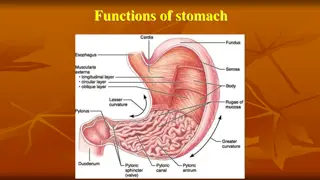
Stomach The stomach is on the left side of the abdominal cavity Cardiac region (near the heart) Fundus (lateral to the cardia) The body (mid and largest portion) Pylorus (funnel-shaped terminal part of the stomach). Most digestive activity occurs in this region. Two sphincters: Cardioesophageal sphincter, through which food enters the stomach from esophagus Pyloric sphincter, which is continuous with small intestine Greater curvature and lesser curvature The lesser omentum (double layer of peritoneum extends from liver to lesser curvature) The greater omentum (another extension of the peritoneum riddled with fat and covers the abdominal organs (cushioning and insulation)
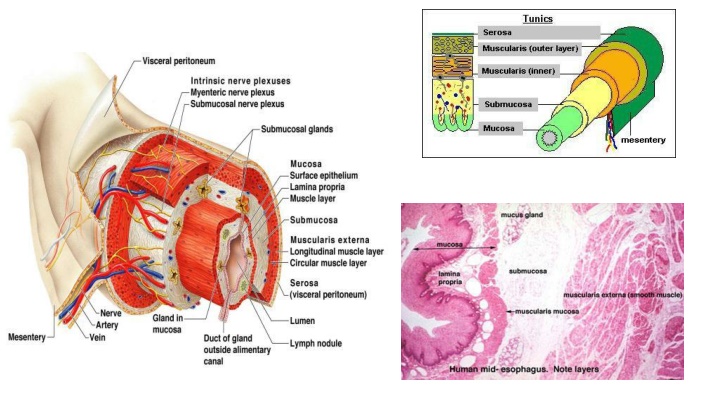
Stomach Histology of the stomach is similar to the esophagus except that the mucosa is lined with simple columnar epithelium which produces large amounts of mucus Digestion: Special arrangement of muscular cells in muscularis externa helps to move, churn and mix the food to break it down to smaller fragments (physical) Chemical breakdown of proteins begins Stomach lining contains deep gastric pits that secrete gastric juice Chief cells produce protein digesting enzymes pepsinogens Parietal cell produce HCL makes stomach acidic and activates enzymes Low PH of stomach stimulates stomach cells to produce gastrin hormone stimulates stomach glands to produce more protein digesting enzymes, mucus and hydrochloric acid How is the stomach wall protected from HCL s corrosive nature? mucous neck cells produce sticky alkaline mucus After food has been processed in the stomach, it resembles heavy cream and is called chime. The chime then enters the small intestine through the pyloric sphincter
Small Intestine The body s major digestive organ Longest section of the alimentary tube (increase surface area for absorption) Extends from pyloric sphincter to ileocecal valve (valve that separates small intestine and large intestine) The large intestine encircles it and frames it in the abdominal cavity Three subdivisions: Duodenum Jejunum (longest part) Ileum (joins large intestine at ileocecal valve)
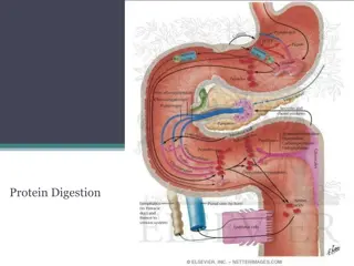
Small Intestine cont. Small intestine can digest only a small amount of food at a time (pyloric sphincter controls food movement from the stomach so that the small intestine is not overwhelmed) Digestion: Most of the digestion occurs in small intestine Enzymes are produced my intestinal cells help in digestion Enzymes produced by pancreas and ducted into the duodenum through the pancreatic ducts Bile (formed by liver) enters duodenum through bile duct Absorption Most of the food absorption occurs in the small intestine
Small Intestine cont. Structures help increase the absorptive surface: Microvilli: tiny projections of the plasma membrane of the mucosal cells, which give the cell surface a fuzzy appearance (brush border) Villi: fingerlike projections of the mucosa. Within each villus is a rich in capillaries and contains a lymphatic capillary called a lacteal Digested food is absorbed through the mucosa cells into the capillaries and lacteal Circular folds, called plicae circulares: deep folds of mucosa and sub-mucosa Peyer s Patches: Collection of lymphatic tissue (high in lymphocytes+ other immune cells) found towards the end of the small intestine (remaining undigested food residue is high in bacteria that must be prevented from entering the blood stream)
Large Intestine Functions Primary task is absorption of water Absorption of other substances (e.g :Bacteria in large intestine that produce vitamin K that is absorbed in the LI) Excretion of wastes Parts of LI: Cecum (where small intestine contents enter the large intestine) Appendix (wormlike structure that hangs from the cecum) Colon Ascending colon (follows the cecum) Transverse colon Descending colon Sigmoid colon (has an S-shaped curve) Rectum Anal canal (opens to the exterior) Has an external voluntary sphincter and internal involuntary sphincter act together to open and close (usually closed except during defecation)
Accessory digestive organs Pancreas: Considered both an Endocrine gland (insulin, glucagon) Exocrine gland (digestive enzymes): trypsinogen that becomes activated into trypsin in the small intestine to digest proteins into amino acids. *Endocrine are ductless glands that secret hormones that travel through the blood to regulate a target organ *Exocrine glands produce secretions that reach their target through a duct Together insulin and glucagon maintain our blood glucose levels insulin lowers blood glucose levels, glucagon increases blood glucose levels
Accessory digestive organs Liver and Gall Bladder: The liver is the largest gland in the body Located under the diaphragm Liver functions include: Helps in digestion by producing bile (yellow to green watery solution containing bile salts,bile pigments, cholesterol and phospholipids) Bile salts helps to break down large fat globules into smaller ones to aid the action of fat-digesting enzymes (lipases) Liver also produces blood clotting factors The gall bladder stores and secretes bile