The Structure and Function of the Heart
Explore the intricate divisions and cell layers of the heart, its chambers and valves, and how it circulates blood throughout the body. Learn about the septa, endocardium, myocardium, epicardium, pericardium, and the vital role of the heart valves in maintaining proper blood flow. Dive deep into the workings of the right and left sides of the heart as they pump blood into different circulations, ensuring oxygenation and proper distribution throughout the body.

Download Presentation

Please find below an Image/Link to download the presentation.
The content on the website is provided AS IS for your information and personal use only. It may not be sold, licensed, or shared on other websites without obtaining consent from the author. If you encounter any issues during the download, it is possible that the publisher has removed the file from their server.
You are allowed to download the files provided on this website for personal or commercial use, subject to the condition that they are used lawfully. All files are the property of their respective owners.
The content on the website is provided AS IS for your information and personal use only. It may not be sold, licensed, or shared on other websites without obtaining consent from the author.
E N D
Presentation Transcript
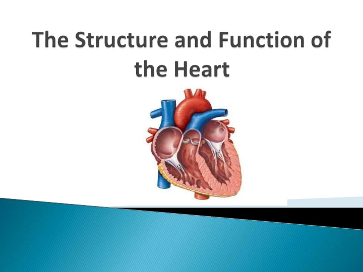
Structure of the Heart The heart is divided into the left and right side by partitions called septa (singular septum). The interatrial septum separates the two upper chambers, called atria (from atri/o, meaning upper chambers ). The interventricular septum separates the two lower chambers, called ventricles (from ventricul/o, meaning lower chamber). Interatrial Septum Interventricular Septum
The Heart Consists of Four Cell Layers: The endocardium (from endo- + cardi/o + -ium, meaning inner layer of the heart ) is formed by endothelial cells, and it lines the interior of the heart chambers and valves. The myocardium (from my/o + cardi/o+ -ium, meaning heart muscle ) is the muscular middle layer of the heart that consists of heart muscle cells. The epicardium (from epi- + cardi/o + -ium, meaning outer layer of the heart ) is formed by epithelial cells, and forms the outer cell layer of the heart. The pericardium (from peri- + cardi/o + -ium, meaning surrounding the heart ) is a membranous sac that surrounds the heart. It consist of two layers called the visceral pericardium (adheres to the epicardium) and parietal pericardium (the outer coat). The space between these two layers is called pericardial cavity and it contains pericardial fluid.
Heart Chambers and Valves The human heart has four chambers, which are responsible for pumping blood and maintaining blood circulation throughout the body. The four chambers are named: The right atrium The left atrium The right ventricle The left ventricle Blood is only pumped to one direction. Four heart valves ensure that blood does not flow backward within the heart.
The Four Heart Valves are Named: The tricuspid valve (from tri- + cuspid, meaning having three points ) located between right atrium andventricle. The pulmonary valve (from pulmon/o, meaning lungs ) located between right ventricle and pulmonary artery. Also called semilunar valve. The mitral valve, also called bicuspid valve ( from bi- + cuspid, meaning having two points ) located between left atrium and ventricle. The aortic valve located between left ventricle and aorta. The tricuspid and bicuspid valves are also called atrioventricular valves (meaning located between the atrium and ventricle ).
Function of the Heart The heart functions to circulate blood aroundthe body. The right and left side of the heart pump blood into two different circulations. The right side pumps deoxygenated (from de- + oxygenated, meaning without oxygen ) blood into the pulmonary circulation, while the left side pumps oxygenated blood into the systemic circulation. The right atrium receives deoxygenated blood from the body tissues via the superior (from super- meaning above ) and inferior (meaning below) vena cava (from ven/o meaning vein ). The blood enters the right atrium, which pumps the blood into the right ventricle. The tricuspid valve prevents blood from flowing backward into the right atrium. The right ventricle pumps the blood into the pulmonary artery via the pulmonary valve.
The pulmonary artery will deliver the deoxygenated blood to the lungs, where gas exchange occurs. Oxygen is taken from the air into the blood (now called oxygenated blood), while carbon dioxide is expelled from the blood into the air. The oxygenated blood returns to the left side of the heart via the pulmonary veins. The oxygenated blood enters the left atrium. The left atrium pumps blood into the left ventricle. The mitral valve prevents blood from flowing backwardinto the left atrium. The left ventricle pumps the blood into the aorta and systemic circulation. The oxygenated blood is delivered everywhere in the body (besides the lungs).
Blood Circulation Blood circulates around the body via two distinct pathways; the pulmonary circulation and the systemic circulation. Together they create a closed pathways that keep the deoxygenated and oxygenated blood separated.
Pulmonary Circulation Pulmonary circulation begins at the right ventricle, where the deoxygenated blood from the body tissues is pumped into the pulmonary arteries and to the lungs. In the lungs, the blood exchanges carbon dioxide (waste product of cellular respiration) to oxygen. The oxygenated blood them travels back to the heart and the left atrium, via the pulmonary vein.
SystemicCircuit The systemic circulation begins at the left ventricle that pumps oxygenated blood into the aorta. Aorta branches out into smaller arteries, which carry the oxygenated blood to the rest of the body (with the exception of lungs). Oxygen is delivered to the body tissues and exchanged to carbon dioxide. The now deoxygenated blood is carried back to the heart and the right atrium via veins.
Arteriesvs Veins The blood vessels that carry blood AWAYfrom heart are called arteries. The bloodvessels that carry blood TOWARD the heart are called veins. Only in systemic circulation arteries carry oxygenated blood, while in the pulmonary circulation arteries carry deoxygenated blood.
Contraction of the Heart The contraction of the muscular wall of the heart chambers, called myocardium generates the force to pump blood. The heart contraction is divided into two phases: systole (meaning contraction ) and diastole (meaning relaxation ). Blood is pumped from the chambers during a contraction phase. The heart chambers are filled with blood during a relaxation phase.
One round of heart contractions can be divided into the following phases: Relaxation phase blood flows from the atria into the ventricles passively via open atrioventricular valves. The atrial systole contraction of atria. Pumps the rest of the blood into the ventricles. The ventricular systole contraction of the ventricles. Forces blood into the pulmonary and systemic circulation. (During the ventricular systole, the atria relax and begin to fill with blood arriving from vena cavaor the pulmonary veins. Ventricular diastole the ventricles and atria are relaxed.
Conduction System of the Heart The conduction system of the heart controls the rate and pattern of your heartbeat.
Sinoatrial (SA) Node Myocardiumcontracts after it receives an electrical impulse generated by a specialized tissue located within the right atrium. This is called the sinoatrialnode (SA node), also called the pacemaker of the heart. The SA node is a bundle of neurons that triggers the contraction of the atria during the cardiac cycle. The electrical currents next reach the ventricles, which contract after the atria. The SA node initiates approximately 75 electrical impulses each minute, with variation between individuals age and general health.
The PurkinjeFibers The Purkinjefibers are cells in the inner ventricle walls, just beneath the endocardium.These fibers run between the ventricles to the apex (bottom) of the heart. The Purkinje fibers play a crucial role in the cardiac cycle. When an electrical stimulus leaves the AVnode,it travels via the bundle of His and branches to the Purkinje fibers. These fibers then carry the impulse throughthe inner wall of each ventricle. This causes the ventricles to contract after the atria contract. The ventricle contractionforces bloodfrom the right ventricle to the lungs (pulmonary circulation) and from the left ventricle to the body (systemic circulation). These three elements generate a healthy heart rhythmknown as sinus rhythm. The rhythm,or contraction of the heart pumps bloodthroughout the body.In roughlya minute s time, bloodtravels from the heart to the bodyand back.
The Electrical Activity of the Heart The electrical activity in the heart is displayed as a P wave, QRS interval and T wave. The P wave correlates to atrial depolarization (systole) and atrial contractions. There is not a wave associated with atrial repolarization as it occurs during ventricular depolarization (during the QRS interval). The QRS complex correlates to ventricular depolarization (systole) as the ventricles contract. The Q wave is the beginning, the R wave the middle of the contraction,and the S wave is the end of ventricular depolarization, and beginning of ventricular repolarization (diastole). The T wave correlates to ventricular repolarization (diastole).
Health Conditions Hypertension: Hypertension is an abnormal condition that is primarily caused by high blood cholesterol. arterialwalls as plaques. These plaques make the lumen of the artery narrower, which causes the blood to flow with higher pressure. Excess cholesterol is deposited on the If an artery becomes completely blocked, the cells supported by that artery will suffer from lack of oxygen and die. If this happens in the coronary arteries, which provide blood to the heart, the result can be myocardial infarction (heart attack).
Stroke: An artery leading to the brain can become blocked. This can cause a cerebral vascular accident, known as a stroke.
Hypotension: Hypotension is also an abnormal condition, in which the blood flows with low pressure. Hypotension occurs when a large volume of water or blood is lost from the body. The body s loss of water, dehydration, can occur during diarrhea or vomiting. The body s loss of blood, hemorrhage, can occur due to blood disorders or injury to the blood vessels (trauma). Hypotension can result in shock.


 Node")


