Thoracentesis Procedure: Indications, Contraindications, Risks, and Precautions
Thoracentesis is a medical procedure used to remove fluid from the pleural space between the lungs and chest wall. This article covers indications, contraindications, risks, and precautions associated with thoracentesis, along with steps to be taken before, during, and after the procedure.

Uploaded on | 0 Views
Download Presentation

Please find below an Image/Link to download the presentation.
The content on the website is provided AS IS for your information and personal use only. It may not be sold, licensed, or shared on other websites without obtaining consent from the author. If you encounter any issues during the download, it is possible that the publisher has removed the file from their server.
You are allowed to download the files provided on this website for personal or commercial use, subject to the condition that they are used lawfully. All files are the property of their respective owners.
The content on the website is provided AS IS for your information and personal use only. It may not be sold, licensed, or shared on other websites without obtaining consent from the author.
E N D
Presentation Transcript
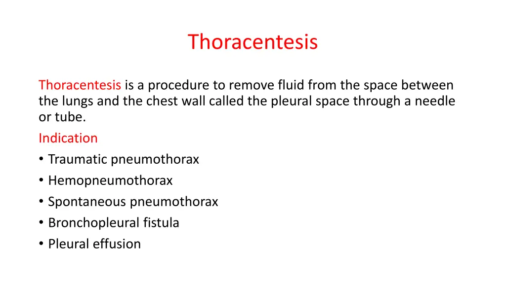
Thoracentesis Thoracentesis is a procedure to remove fluid from the space between the lungs and the chest wall called the pleural space through a needle or tube. Indication Traumatic pneumothorax Hemopneumothorax Spontaneous pneumothorax Bronchopleural fistula Pleural effusion
Contraindication Contraindication Coagulation disorder Atelectasis Emphysema(pulmonary enlargement) Severe cough or hiccups COMPLICATION Pulmonary edema Bleeding Respiratory distress Infection Air embolism Dyspnea and cough Cardiac tamponade(fluid build up in the space between myocardium and pericardium ) Atelectasis (lung collapse)
Before The Procedure Explain the purpose, risks/benefits, and steps of the procedure and obtain consent from the patient or appropriate legal design. R: An explanation helps orient the patient to the procedure assist in coping and provide an opportunity to ask question and verbalise anxiety Take Medical History such as Trouble in breathing, coughing, and hiccups, Had heart disease, Smoked Prepare Equipment Dressing set Povidone / Alcohol Abraham s needle Local anaesthetic, e.g. lignocaine (lidocaine) 1% Connecting tubing or 2% Syringe 50ml and 5ml Formalin bottle Urine bottle x2 Needles (18 and 23 C+S bottle gauge) 3-way stopcock Sterile Glove
Place patient upright / cardiac position and help patient maintain position during procedure. R: the upright position ensures that the diaphragm is more dependent and facilitates the removal of fluid that usually localizes at the base of the chest. Explain that he/she will receive a local anesthetic to minimize pain during the procedure. Clean patient skin with antiseptic to prevent infection and maintain
DURING PROCEDURE Observe patient respiration rate and breathing pattern Assess patient vital sign to prevent any complication such as hypovolemic shock during procedure. Observe patient level of consciousness and give emotional support to reduce patient anxiety Monitor saturation to prevent hypoxia. AFTER PROCEDURE Obtain a chest x-ray to evaluate the fluid level To compare the conditions of the lungs before and after the procedure. Document the procedure, patient s response, characteristics of fluid and amount, and patient response to follow-up Provide post-procedural analgesics as needed To prevent patient from pain related to the incision site
Paracentesis A sterile procedure to remove fluid (ascites) from the peritoneal cavity. Indications Diagnostic: Unexplained ascites (e.g., infection, cancer). Suspected spontaneous bacterial peritonitis (SBP). Therapeutic: Respiratory distress due to fluid pressure. Abdominal pain/discomfort.
Contraindications Severe bleeding disorders. Bowel obstruction. Skin infection at puncture site. Equipment Needed Sterile tray: Needle,syringes, tubes. Dressing supplies: Gauze, adhesive bandage. Lab tubes: Culture, albumin, cell count. IV access kit (for fluid replacement if needed).
Procedure Nursing Care 1.Patient Education: Explain steps, obtain consent. 2.Assessment: 1.Vital signs (baseline). 2.Abdominal girth, weight. 3.Preparation: 1.Empty bladder. 2.Position: Supine or slightly lateral. 4.Site: Midline or lateral lower quadrant (ultrasound-guided). 5.Aseptic technique: Chlorhexidine prep, sterile drapes. 6.Fluid removal: 1.Diagnostic: 20 50 mL. 2.Therapeutic: Up to 4 6 L (monitor for hypotension).
Complications Hypotension, mild bruising. Infection (peritonitis). Bowel perforation. Hemorrhage Patient Education Report fever, dizziness, or abdominal pain. Limit activity for 24 hours. Follow-up for lab results (if diagnostic).