Thyroglossal Duct Cysts and Neck Masses
Thyroglossal duct cysts are common congenital neck masses, presenting as midline enlarging masses in young individuals. This overview covers their embryological development, clinical features, diagnostic imaging, and management.

Download Presentation

Please find below an Image/Link to download the presentation.
The content on the website is provided AS IS for your information and personal use only. It may not be sold, licensed, or shared on other websites without obtaining consent from the author.If you encounter any issues during the download, it is possible that the publisher has removed the file from their server.
You are allowed to download the files provided on this website for personal or commercial use, subject to the condition that they are used lawfully. All files are the property of their respective owners.
The content on the website is provided AS IS for your information and personal use only. It may not be sold, licensed, or shared on other websites without obtaining consent from the author.
E N D
Presentation Transcript
DR. Kavita Makasare JR III
Usually the 1stinvestigation. Relationship to the surrounding structures. Confirms the cystic nature of lesion.
For evaluation of soft tissue planes adjacent to large masses that cannot be visualized on USG. Superior for detection of calcification. When contrast is used, tells us about the vascularity of the lesion.
Multiplanar capability Superior contrast resolution. Especially useful in cases with extension into mediastinum or deep spaces of the neck. Superior resolution for masses located in anatomically complex areas such as the floor of mouth.
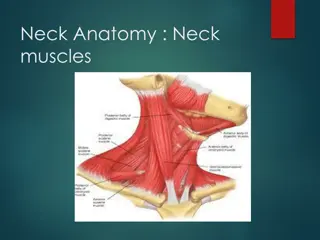
Development : in the 3rdwk of foetal life : foramen caecum : the junction of the anterior 2/3rdand posterior 1/3rdof tongue. Descends and penetrates floor of mouth and tongue musculature Anterior to hyoid bone and laryngeal cartilage Final position :7thwk : descending anterior to the thyrohyoid membrane and strap muscles of the neck.
During its migration : the tract forms the thyroglossal duct : involutes by 8th-10th week. If any part persists : secretions from the lining epithelium forms the thyroglossal cyst. Site of original opening : At tongue : foramen caecum Inferior end : pyramidal lobe of thyroid.
MC congenital neck mass (70%). 2ndMC neck mass (second to benign cervical lymphadenopathy). M=F (no gender predilection). Rarely hereditary : AD : prepubertal females.
MC located in midline (75%) or within 2cms of midline (25%). If off midline, mostly on left for reasons not understood. 80% are seen at or below the level of hyoid bone. Enlarging painless mass which moves upwards with protrusion of tongue in a young pt.
In 1% cases, from the ectopic rests of thyroid tissue within it and not from the duct itself. MC : papillary type (80%), but virtually all types have been reported. Nodal spread is less common as compared to primary carcinoma arising in the thyroid gland itself.
CHARACTERISTIC location : cyst like mass in the anterior neck in the midline at the level of hyoid bone or within the strap muscles. Anechoic mass in the characteristic location : CLASSIC APPEARANCE : less common. More common : hypoechoic lesion (homogenous/ heterogenous) with increased through transmission : with internal echos.
Presence/ absence of internal echos does not correlate with the pathological presence of inflammation. Mostly due to the presence of proteinaceous material within the cyst. Preoperative visualization of the normal thyroid tissue excludes the presence of ectopic thyroid tissue.
Smooth well circumscribed mass anywhere along the course of the duct. Thin walled, with peripheral rim enhancement on post contrast study. Homogenous attenuation (10-18HU). Increased attenuation suggest increased protein content and correlated with prior history of infection. Usually unilocular, occasionally septations+ Beaking of strap muscles over the surface of cyst.
CECT shows a cystic lesion in the right strap muscles.
UNCOMPLICATED CYST : hypo on T1W and hyper on T2WI with smooth non enhancing wall. INFECTED/HAEMORRHAGIC CYST: thick irregular wall with enhancement. variable signal intensities depending on the protein content.
Simple incision and drainage or partial resection almost always gives rise to recurrence. Complete excision: SISTRUNK PROCEDURE : involves excision of a central portion of the hyoid bone and a core of tissue following the expected course of thyroglossal duct to the foramen caecum. Recurrence rate : 2.6%
1starch : Mandibular arch. 2ndarch : Hyoid arch 3rd, 4thand 6tharches donot have special names. 5tharch disappears soon after its formation.
1STcleft : dorsal part : develops into the epithelial lining of the EAC. Ventral part obliterates. 2nd, 3rdand 4thclefts form a part of the ectodermally lined depression known as the Cervical Sinus of His.
1STpouch : ventral part- tongue Dorsal part with dorsal part of 2ndforms a diverticulum called as tubotympanic recess Forms the eustachian tube, middle ear cavity and tympanic antrum. 2ndpouch : ventral part : tonsils 3rdpouch: inferior parathyroid gland and thymus. 4thpouch : superior parathyroid gland and part of thyroid.
Branchial cleft anomalies arise from incomplete obliteration of the Cervical Sinus of His or from epithelial cell rests. Manifest as sinus, fistula or cysts. Patients presenting as cyst : older children or young adults. fistulas: infants or young children. Bilateral in 2-3%
Also known as parotid lymphoepithelial cyst. Uncommon cyst Arise in the 1stbranchial arch tract from the EAC through the parotid gland into the submandibular triangle. MC in middle aged women. C/F : recurrent abscess or sinus around the ear or angle of mandible. Facial nerve palsy may occur.
On CT : cystic lesion either within, superficial or deep to the parotid gland. Wall thickness and enhancement is variable and increases with repeated infections.
95% of the branchial cleft anomalies arise from the 2nd arch. 3/4thof these are cysts (manifest between 10-40yrs) in contrast to sinus or fistulas (manifest in 1st decade). No gender prediliction.
Usually occur in the submandibular space. Present as a painless, fluctuating mass in the lateral portion of the neck at the anteromedial border of the SCM at the mandibular angle. Slowly grows Painful and tender : if recurrent infection.
TYPE I : most superficial Lies along the anterior surface of the SCM just deep to the platysma muscle. TYPE II : MC and found in the MC location of these cysts Along the anterior border of the SCM lateral to the carotid space and posterior to the submandibular gland.
TYPE III : extends medially between the bifurcation of the internal and external carotid artery upto the lateral pharyngeal wall. TYPE IV : lies in the pharyngeal mucosal space and is lined by columnar epithelium.
USG : sharply marginated rounded anechoic mass with a thin wall displacing the surrounding structures with posterior acoustic enhancement. Occasional internal echos due to debris. CT : uniform homogenous hypodense lesion with a uniform thin wall. Mural thickness may increase after infection.
CHARACTERISTIC LOCATION : Along the anterior border of the SCM lateral to the carotid space and posterior to the submandibular gland. Displaces the carotid vessels : medially\ SCM : posteriorly or posterolaterally Submandibular gland : anteriorly May extend into the parapharyngeal space after extending through the stylomandibular tunnel and middle constrictor muscle.
MRI better depicts the deep tissue extent. Cyst fluid hypo/hyper on T1W and hyperintense on T2WI. Occasionally, BEAK SIGN : on axial CT/MRI image : pathognomic of a 2ndbranchial cleft cyst especially bailey s type III cyst It represents a curved rim of tissue or a beak pointing medially between the ECA and ICA.
Extremely rare. 3rdcyst is the 2ndMC lesion in the posterior cervical space after cystic hygroma. Majority of the 4thcleft anomalies manifest as fistulas rather than cyst or sinus.
3rdcleft cyst : Posterior to the common or the internal carotid artery If fistula : pierce the thyrohyoid membrane and reach the pyriform sinus. Most are located in the posterior cervical space, posterior to the SCM. 4th cleft sinus tract : Arise from the pyriform sinus, pierce the thyrohyoid membrane and begins to descend in the mediastinum in the tracheo-oesophageal groove.
3rdcyst : appears as a unilocular cystic mass in the posterior cervical space. Signal intensity varies according to the fluid content. 4thcleft cyst which connect to the pyriform sinus : appear similar to a external or a mixed laryngocele.
Most common form of lymphangioma of infancy and childhood Age : 2 years (80-90%) Gender : No predilection Pathogenesis : Early sequestration of embryonic lymphatic channels, more commonly from developing jugular lymph sac pair.
Site : 75-80% in neck and lower face Children : posterior cervical space> oral cavity Adults : Sublingual, subamandibular, parotid spaces Less common- Axilla, mediastinum, abdominal cavity, retroperitoneum, scrotum, skeleton Infiltrative lesions, do not respect fascial planes
Cystic hygroma Clinical features. Majority painless masses in neck Large masses pressure Sudden increase- hemorrhage/trauma/viral infection Facial nerve palsy, dysphagia, feeding problems Chylothorax/ chylomediastinum mediastinal involvement
US- multilocular, predominantly cystic with variably thick septae. Echogenic areas- cluster of abnormal lymphatic channels. Fluid-fluid levels hemorrhage


 or within 2cms")