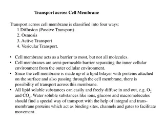
Transport of Small Molecules
The internal composition of a cell is maintained by the selectively permeable plasma membrane, allowing control over molecule exchange. Passive diffusion, mediated by transport proteins, permits small molecules to cross the membrane easily. However, larger polar or charged molecules require specific transport proteins for passage. Learn about the permeability of phospholipid bilayers and the mechanisms by which different molecules move across cell membranes.

Download Presentation

Please find below an Image/Link to download the presentation.
The content on the website is provided AS IS for your information and personal use only. It may not be sold, licensed, or shared on other websites without obtaining consent from the author.If you encounter any issues during the download, it is possible that the publisher has removed the file from their server.
You are allowed to download the files provided on this website for personal or commercial use, subject to the condition that they are used lawfully. All files are the property of their respective owners.
The content on the website is provided AS IS for your information and personal use only. It may not be sold, licensed, or shared on other websites without obtaining consent from the author.
E N D
Presentation Transcript
Transport of Small Molecules The internal composition of the cell is maintained because the plasma membrane is selectively permeable to small molecules. Most biological molecules are unable to diffuse through the phospholipid bilayer, so the plasma membrane forms a barrier that blocks the free exchange of molecules between the cytoplasm and the external environment of the cell. Specific transport proteins (carrier proteins and channel proteins) then mediate the selective passage of small molecules across the membrane, allowing the cell to control the composition of its cytoplasm.
Passive Diffusion The simplest mechanism by which molecules can cross the plasma membrane is passive diffusion. During passive diffusion, a molecule simply dissolves in the phospholipid bilayer, diffuses across it, and then dissolves in the aqueous solution at the other side of the membrane. No membrane proteins are involved and the direction of transport is determined simply by the relative concentrations of the molecule inside and outside of the cell. The net flow of molecules is always down their concentration gradient from a compartment with a high concentration to one with a lower concentration of the molecule.
Passive diffusion is thus a nonselective process by which any molecule able to dissolve in the phospholipid bilayer is able to cross the plasma membrane and equilibrate between the inside and outside of the cell. Importantly, only small, relatively hydrophobic molecules are able to diffuse across a phospholipid bilayer at significant rates. Thus, gases (such as O2and CO2), hydrophobic molecules (such as benzene), and small polar but uncharged molecules (such as H2O and ethanol) are able to diffuse across the plasma membrane.
Other biological molecules are unable to dissolve in the hydrophobic interior of the phospholipid bilayer. Consequently, larger uncharged polar molecules such as glucose are unable to cross the plasma membrane by passive diffusion, as are charged molecules of any size (including small ions such as H+, Na+, K+, and Cl-). The passage of these molecules across the membrane instead requires the activity of specific transport and channel proteins, which therefore control the traffic of most biological molecules into and out of the cell.
Gases, hydrophobic molecules, and small polar uncharged molecules can diffuse through phospholipid bilayers. Larger polar molecules and charged molecules cannot. Permeability of phospholipid bilayers
Facilitated Diffusion and Carrier Proteins Facilitated diffusion, like passive diffusion, involves the movement of molecules in the direction determined by their relative concentrations inside and outside of the cell. No external source of energy is provided, so molecules travel across the membrane in the direction determined by their concentration gradients and, in the case of charged molecules, by the electric potential across the membrane. However, facilitated diffusion differs from passive diffusion in that the transported molecules do not dissolve in the phospholipid bilayer.
Instead, their passage is mediated by proteins that enable the transported molecules to cross the membrane without directly interacting with its hydrophobic interior. Facilitated diffusion therefore allows polar and charged molecules, such as carbohydrates, amino acids, nucleosides, and ions, to cross the plasma membrane.
Two classes of proteins that mediate facilitated diffusion are generally distinguished: carrier proteins and channel proteins. Carrier proteins bind specific molecules to be transported on one side of the membrane. They then undergo conformational changes that allow the molecule to pass through the membrane and be released on the other side. Channel proteins form open pores through the membrane, allowing the free diffusion of any molecule of the appropriate size and charge.
Carrier proteins are responsible for the facilitated diffusion of sugars, amino acids, and nucleosides across the plasma membranes of most cells. The uptake of glucose, which serves as a primary source of metabolic energy and the glucose transporter provides a well-studied example of a carrier protein. The glucose transporter was initially identified as a 55-kd protein in human red blood cells, in which it represents approximately 5% of total membrane protein.
Subsequent isolation and sequence analysis of a cDNA clone revealed that the glucose transporter has 12 -helical transmembrane segments a structure typical of many carrier proteins. These transmembrane helices contain predominantly hydrophobic amino acids, but several also contain polar amino acid residues that are thought to form the glucose-binding site in the interior of the protein.
The glucose transporter has 12 transmembrane helices. Polar amino acid residues located within the phospholipid bilayer are indicated as dark purple circles. Structure of the glucose transporter
As with many membrane proteins, the three-dimensional structure of the glucose transporter is not known. Kinetic studies indicate that the glucose transporter functions by alternating between two conformational states. In the first conformation, a glucose-binding site faces the outside of the cell. The binding of glucose to this exterior site induces a conformational change in the transporter, such that the glucose-binding site now faces the interior of the cell. Glucose can then be released into the cytosol, followed by the return of the transporter to its original conformation.
The glucose transporter alternates between two conformations in which a glucose- binding site is alternately exposed on the outside and the inside of the cell. In the first conformation shown (A), glucose binds to a site exposed on the outside of the plasma membrane. The transporter then undergoes a conformational change such that the glucose-binding site faces the inside of the cell and glucose is released into the cytosol (B). The transporter then returns to its original Model for the facilitated diffusion of glucose conformation (C).
Most cells, including erythrocytes, are exposed to extracellular glucose concentrations that are higher than those inside the cell, so facilitated diffusion results in the net inward transport of glucose. Once glucose is taken up by these cells it is rapidly metabolized, so intracellular glucose concentrations remain low and glucose continues to be transported into the cell from the extracellular fluids. Because the conformational changes of the glucose transporter are reversible, however, glucose can be transported in the opposite direction simply by reversing the steps. Such reverse flow occurs in liver cells, in which glucose is synthesized and released into the circulation.
Ion Channels In contrast to carrier proteins, channel proteins simply form open pores in the membrane, allowing small molecules of the appropriate size and charge to pass freely through the lipid bilayer. One group of channel proteins is the porins, which permit the free passage of ions and small polar molecules through the outer membranes of bacteria. Channel proteins also permit the passage of molecules between cells connected at gap junctions.
The plasma membranes of many cells also contain water channel proteins (aquaporins), through which water molecules are able to cross the membrane much more rapidly than they can diffuse through the phospholipid bilayer. The best-characterized channel proteins, however, are the ion channels, which mediate the passage of ions across plasma membranes. Although ion channels are present in the membranes of all cells, they have been especially well studied in nerve and muscle, where their regulated opening and closing is responsible for the transmission of electric signals.
Three properties of ion channels are central to their function. First, transport through channels is extremely rapid. More than a million ions per second flow through open channels a flow rate approximately a thousand times greater than the rate of transport by carrier proteins. Second, ion channels are highly selective because narrow pores in the channel restrict passage to ions of the appropriate size and charge.
Thus, specific channel proteins allow the passage of Na+, K+, Ca2+, and Cl- across the membrane. Third, most ion channels are not permanently open. Instead, the opening of ion channels is regulated by gates that transiently open in response to specific stimuli. Some channels (called ligand-gated channels) open in response to the binding of neurotransmitters or other signaling molecules. Others (voltage-gated channels) open in response to changes in electric potential across the plasma membrane.
In the closed conformation, the flow of ions is blocked by a gate. Opening of the gate allows ions to flow rapidly through the channel. The channel contains a narrow pore that restricts passage to ions of the appropriate size and charge. Model of an ion channel
The fundamental role of ion channels in the transmission of electric impulses was elucidated through a series of elegant experiments reported by Alan Hodgkin and Andrew Huxley in 1952. These investigators used the giant nerve cells of the squid as a model. The axons of these giant neurons have a diameter of about 1 mm, making it possible to insert electrodes and measure the changes in membrane potential that take place during the transmission of nerve impulses.
Using this approach, Hodgkin and Huxley demonstrated that these changes in membrane potential result from the regulated opening and closing of Na+ and K+ channels in the plasma membrane. It subsequently became possible to study the activity of individual ion channels, using the patch clamp technique developed by Erwin Neher and Bert Sakmann in 1976. In this method, a micropipette with a tip diameter of about 1 m is used to isolate a small patch of membrane, allowing the flow of ions through a single channel to be analyzed and greatly increasing the precision with which the activities of ion channels can be studied.
A small patch of membrane is isolated in the tip of a micropipette. Stimuli can be applied from within the pipette, allowing the behaviour of the trapped channel to be measured. The patch clamp technique
The flow of ions through membrane channels is dependent on the establishment of ion gradients across the plasma membrane. All cells, including nerve and muscle, contain ion pumps that use energy derived from ATP hydrolysis to actively transport ions across the plasma membrane. As a result, the ionic composition of the cytoplasm is substantially different from that of extracellular fluids. For example, Na+ is actively pumped out of cells while K+ is pumped in. In the squid axon the concentration of Na+ is about 10 times higher in extracellular fluids than inside the cell, whereas the concentration of K+ is approximately 20 times higher in the cytosol than in the surrounding medium.
Because ions are electrically charged, their transport results in the establishment of an electric gradient across the plasma membrane. With resting squid axons there is an electric potential of about 60 mV across the plasma membrane, with the inside of the cell negative with respect to the outside. This electric potential arises both from ion pumps and from the flow of ions through channels that are open in the resting cell plasma membrane. The plasma membrane of resting squid axons contains open K+ channels, so it is more permeable to K+ than to Na+ or other ions. Consequently, the flow of K+ makes the largest contribution to the resting membrane potential.
Ion gradients and resting membrane potential of the giant squid axon Only the concentrations of Na+ and K+ are shown, because these are the ions that function in the transmission of nerve impulses. Na+ is pumped out of the cell while K+ is pumped in, so the concentration of Na+ is higher outside than inside of the axon, whereas the concentration of K+ is higher inside than out. The resting membrane is more permeable to K+ than to Na+ or other ions because it contains open K+ channels. The flow of K+ through these channels makes the major contribution to the resting membrane potential of -60 mV, which is therefore close to the K+ equilibrium potential.
The flow of ions across a membrane is driven by both the concentration and voltage components of an electrochemical gradient. For example, the 20-fold higher concentration of K+ inside the squid axon as compared to the extracellular fluid drives the flow of K+ out of the cell. However, because K+ is positively charged, this efflux of K+ from the cell generates an electric potential across the membrane, with the inside of the cell becoming negatively charged. This membrane potential opposes the continuing flow of K+ out of the cell, and the system approaches the equilibrium state, in which the membrane potential balances the K+ concentration gradient. Quantitatively, the relationship between ion concentration and membrane potential is given by the Nernst equation:
where V is the equilibrium potential in volts, R is the gas constant, T is the absolute temperature, z is the charge of the ion, F is Faraday's constant, and Co and Ci are the concentrations of the ion outside and inside of the cell, respectively. An equilibrium potential exists separately for each ion, and the membrane potential is determined by the flow of all the ions that cross the plasma membrane. However, because resting squid axons are more permeable to K+ than to Na+ or other ions (including Cl-), the resting membrane potential (-60 mV) is close to the equilibrium potential determined by the intracellular and extracellular K+ concentrations (-75 mV).
As nerve impulses (action potentials) travel along axons, the membrane depolarizes. The membrane potential changes from -60 mV to approximately +30 mV in less than a millisecond, after which it becomes negative again and returns to its resting value. These changes result from the rapid sequential opening and closing of voltage- gated Na+ and K+ channels. Relatively small initial changes in membrane potential (from -60 to about -40 mV) lead to the rapid opening of Na+ channels. This allows Na+ to flow into the cell, driven by both its concentration gradient and the membrane potential.
The sudden entry of Na+ leads to a large change in membrane potential, which increases to nearly +30 mV, approaching the Na+ equilibrium potential of approximately +50 mV. At this time, the Na+ channels are inactivated and voltage-gated K+ channels open, substantially increasing the permeability of the membrane to K+. K+ then flows rapidly out of the cell, driven by both the membrane potential and the K+ concentration gradient, leading to a rapid decrease in membrane potential to about -75 mV (the K+ equilibrium potential). The voltage-gated K+ channels are then inactivated and the membrane potential returns to its resting level of -60 mV, determined by the flow of K+ and other ions through the channels that remain open in unstimulated cells.
Membrane potential and ion channels during an action potential (A) Changes in membrane potential at one point on a squid giant axon following a stimulus. ENa and EK are the equilibrium potentials for Na+ and K+, respectively. (B) The membrane potential first increases as voltage-gated Na+ channels open. The membrane potential then falls below its resting value as the Na+ channels are inactivated and voltage-gated K+ channels open. The voltage-gated K+ channels are then inactivated, and the membrane potential returns to its resting value.
Depolarization of adjacent regions of the plasma membrane allows action potentials to travel down the length of nerve cell axons as electric signals, resulting in the rapid transmission of nerve impulses over long distances. For example, the axons of human motor neurons can be more than a meter long. The arrival of action potentials at the terminus of most neurons then signals the release of neurotransmitters, such as acetylcholine, which carry signals between cells at a synapse. Neurotransmitters released from presynaptic cells bind to receptors on the membranes of postsynaptic cells, where they act to open ligand-gated ion channels.
One of the best-characterized of these channels is the acetylcholine receptor of muscle cells. Binding of acetylcholine opens a channel that is permeable to both Na+ and K+. This permits the rapid influx of Na+, which depolarizes the muscle cell membrane and triggers an action potential. The action potential then results in the opening of voltage-gated Ca2+ channels, leading to the increase in intracellular Ca2+ that signals contraction.
Signaling by neurotransmitter release at a synapse The arrival of a nerve impulse at the terminus of the neuron signals the fusion of synaptic vesicles with the plasma membrane, resulting in the release of neurotransmitter from the presynaptic cell into the synaptic cleft. The neurotransmitter binds to receptors and opens ligand-gated ion channels in the target cell plasma membrane.
The acetylcholine receptor, initially isolated from the electric organ of Torpedo rays in the 1970s, is the prototype of ligand-gated channels. The receptor consists of five subunits arranged as a cylinder in the membrane. In its closed state, the channel pore is thought to be blocked by the side chains of hydrophobic amino acids. The binding of acetylcholine induces a conformational change in the receptor such that these hydrophobic side chains shift out of the channel, opening a pore that allows the passage of positively charged ions, including Na+ and K+. However, the channel remains impermeable to negatively charged ions, such as Cl-, because it is lined by negatively charged amino acids.
Model of the acetylcholine receptor The receptor consists of five subunits arranged around a central pore. The binding of acetylcholine to a site in the extracellular region of the receptor induces allosteric changes that open the channel gate. The channel is lined by negatively charged amino acids that prevent the flow of negatively charged ions.
A greater degree of ion selectivity is displayed by the voltage-gated Na+ and K+ channels. Na+ channels are more than ten times more permeable to Na+ than to K+, whereas K+ channels are more than a thousand times more permeable to K+ than to Na+. The selectivity of the Na+ channel can be explained, at least in part, on the basis of a narrow pore that acts as a size filter. The ionic radius of Na+ (0.95 ) is smaller than that of K+ (1.33 ), and it is thought that the Na+ channel pore is narrow enough to interfere with the passage of K+ or larger ions.
Ion selectivity of Na+channels A narrow pore permits the passage of Na+ bound to a single water molecule but interferes with the passage of K+ or larger ions.
K+ channels also have narrow pores, which prevent the passage of larger ions. However, since Na+ has a smaller ionic radius, this does not account for the selective permeability of these channels to K+. Selectivity of the K+ channel is based on a different mechanism, which was elucidated with the determination of the three-dimensional structure of a K+ channel by X-ray crystallography in 1998. The channel pore contains a narrow selectivity filter that is lined with carbonyl oxygen (C=O) atoms from the polypeptide backbone.
When a K+ ion enters the selectivity filter, interactions with these carbonyl oxygens displace the water molecules to which K+ is bound, allowing dehydrated K+ to pass through the pore. In contrast, a dehydrated Na+ is too small to interact with these carbonyl oxygens in the selectivity filter, which is held rigidly open. Consequently, Na+ remains bound to water molecules in a hydrated complex that is too large to pass through the channel.
Selectivity of K+channels The K+ channel contains a narrow selectivity filter lined with carbonyl oxygen (C=O) atoms. The pore is just wide enough to allow the passage of dehydrated K+ from which all associated water molecules have been displaced as a result of interactions between K+ and these carbonyl oxygens. Na+ is too small to interact with the carbonyl oxygens of the selectivity filter, so it remains bound to water in a complex that is too large to pass through the channel pore.
Voltage-gated Na+, K+, and Ca2+ channels all belong to a large family of related proteins. For example, the genome sequence of C. elegans has revealed nearly 200 genes encoding ion channels, which presumably are needed to play diverse roles in cell signaling. K+ channels consist of four identical subunits, each containing either two or six transmembrane helices. Na+ and Ca2+ channels consist of a single polypeptide chain, but each polypeptide contains four repeated domains that correspond to the K+ channel subunits.
Voltage gating is mediated by one of the transmembrane helices, which contains multiple positively charged amino acids. Membrane depolarization induces the movement of these positive charges toward the outside of the cell, shifting the position of this transmembrane segment and opening the channel. Rapid inactivation of Na+ and K+ channels during the propagation of action potentials is then mediated by cytoplasmic portions of the polypeptide chain, which bind to the cytoplasmic mouth of the channel pore and prevent further ion flow.
Structures of voltage-gated cation channels The K+, Na+, and Ca2+ channels belong to a family of related proteins. The K+ channel is formed from the association of four identical subunits, one of which is shown. The Na+ channel consists of a single polypeptide chain containing four repeated domains, each of which is similar to one K+ channel subunit. The Ca2+ channel is similar to the Na+ channel. Each subunit or domain contains six membrane-spanning helices. The helix designated 4 contains multiple positively charged amino acids and acts as the voltage sensor that mediates channel opening in response to changes in membrane potential.
Inactivation of K+and Na+channels Following voltage-gated opening, the K+ and Na+ channels are rapidly inactivated by the binding of cytoplasmic portions of the polypeptide chains to the pore. For the K+ channel, inactivation is mediated by a ball-and-chain mechanism with the ball corresponding to the amino terminus of the polypeptide chain. For the Na+ channel, inactivation is mediated by the intracellular loop connecting domains III and IV.
A wide variety of ion channels (including Ca2+ and Cl- channels) respond to different neurotransmitters or open and close with different kinetics following membrane depolarization. The concerted actions of these multiple channels are responsible for the complexities of signaling in the nervous system. Moreover, as discussed in the next chapter, the roles of ion channels are not restricted to the electrically excitable cells of nerve and muscle; they also play critical roles in signaling in other cell types. The regulated opening and closing of ion channels thus provides cells with a sensitive and versatile mechanism for responding to a variety of environmental stimuli.