Trichomonas Vaginitis in Humans
Trichomonas vaginalis is a pathogenic parasite causing vaginitis in humans. This parasitic infection can lead to various clinical symptoms such as foul-smelling discharge, itching, and urethral involvement. The life cycle of Trichomonas species involves trophozoites residing in the mucosal surfaces and feeding on local bacteria and leukocytes.

Download Presentation

Please find below an Image/Link to download the presentation.
The content on the website is provided AS IS for your information and personal use only. It may not be sold, licensed, or shared on other websites without obtaining consent from the author.If you encounter any issues during the download, it is possible that the publisher has removed the file from their server.
You are allowed to download the files provided on this website for personal or commercial use, subject to the condition that they are used lawfully. All files are the property of their respective owners.
The content on the website is provided AS IS for your information and personal use only. It may not be sold, licensed, or shared on other websites without obtaining consent from the author.
E N D
Presentation Transcript
Lab 2:- Parasitology asst.l. Mohammed Abdulrasool
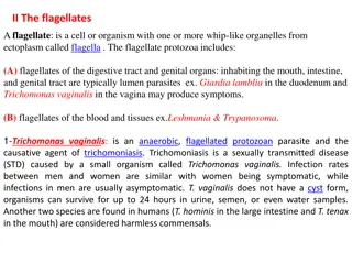
2.Trichomonas vaginalis 1. Pathogenic to human &causes vaginitis (trichomoniasis). 2. troph. Is round or pear like in shape, contains 4-6 flagella, all originating from anterior end & only one extend posteriorly. The motility is rapid & jerky. The undulating membrane extending half of the body length. Prominent axostyle that often curves around the nucleus & granules may be seen along in the axostyle. The nucleus is oval shape & only one. No cyst is seen. Clinical symptoms 1. T. vaginalis reside on the mucosal surface of the vagina in infected women. The most common sites in male is the prostate gland region & the epithelium of the urethra. 2. Vaginitis may be found in infected women. It is characterized by foul smelling, greenish- 3. yellow, vaginal discharge, burning &itching may also present. Red lesions may be seen in vaginal mucosa. Urethral involvement, dysuria & increased frequency of urination are among the most commonly symptoms. Cystitis is rare occur.
Trichomonas vaginalis Trichomonas tenax
Life cycle Trichomonas vaginalis trophozoites reside on the mucosal surface of the vagina in infected women. The growing trophozoites multiply by longitudinal binary fission and feed on local bacteria and leukocytes. The Trichomonas vaginalis trophozoites thrive in a slightly alkaline or slightly acid PH environment, such as that commonly seen in an unhealthy vagina. The most common infection site of T. vaginalis in males is the prostate gland region and the epithelium of the urethra. The detailed life cycle in the male host is unknown. Infective and diagnostic stage is trophozoites
3.Trichomonas tenax Trophzoite: Oval to pear in Shape. Have one nuclei, vesicular filled with chromatin granules. Have five flagella, all originating anteriorly, four extends anteriorly, one extends posteriorly. Undulating membrane extending 2/3 of body length. Thick axostyle and Small anterior cytosome opposite undulating membrane. There is a known cyst Life cycle Trichomonas tenax trophozoites survive in the body as mouth scavengers that feed primarily on local microorganisms. Located in the tartar between the teeth, tonsillar crypts pyorrheal pockets, and gingival margin around the gums, T. tenax trophozoites multiply by longitudinal binary fission. These trophozoites are unable to survive the digestive process.
Clinical symptoms The typical Trichomonas tenax infection does not produce any notable symptoms. On a rare occasion, T. tenax has been known to invade the respiratory tract, but this appears to have mainly occurred in patients with underlying thoracic or lung abscesses of pleural exudates.
B:Class Haemoflagellates: Leishmania & Trypanosoma 1. Blood and Tissue Protozoal Infections The major protozoal diseases that involve the blood and internal organs are malaria (Plasmodium species), toxoplasmosis (Toxoplasma species), trypanosomiasis (Trypanosoma species), and leishmaniasis (Leishmania species). Plasmodium and Toxoplasma are sporozoans, whereas Trypanosoma and Leishmania are flagellates, sometimes referred to as hemoflagellates. These parasites are unicellular with flagellum in the beginning of the parasite which helping it in motile. Leishmania = mediated host= Sand fly Trypanosoma= mediated host= Tse-Tse fly
1. These parasites invading the blood, the tissues, and endothelial layer of organs and the tissues of skin. 2. These flagellates including two genus that important to human: Leishmania: This genus is circular or ovum in shape with one nucleus located in the center of the cell and in front of the nucleus present the motile generator which is short. Trypanosoma: The cell of this genus is tall with one central nucleus and the motile generator located in the terminal of parasite. The flagellate belong to these two genus pass through the life cycle; the live cycle .1 between two host: vertebrate host(terminal host like human)and arthropod host (mediated host like the fly) in many stages with different shapes, through the shape of the body, presence of flagellate or absent, the shape locate of motile generator and the presence of waved membrane or absent ,as following:
1.: A. Amastigote: the parasite circular or ovum in shape,the nucleus lies near the center and it in front of it present motile generator which extend short flagella from it and have not waved membrane. B. Promastigote: thebody is spindle with nucleus in the center and motile generator located near the beginning of the body arise a flagellate from generator extend out of the body and have not waved membrane.
C. Epimastigote: the body is tall and motile generator lies in front of the nucleus that move little away from the center of the body and arise on it flagellate that connect with the body and with the waved membrane therefore extend with free end.
D. Trypomastigote:Thebody is spindle and the nucleus lies in the center,the motile generator lies in the last part of the parasite and arise from it a flagellate extend along the external adage to the waved membrane and the end of flagella is free. Note:the parasite that belong to Leishmania pass through the life cycle in amastigote and promastigote forms while Trypanosoma pass through the life cycle with all forms.
Leishmania: Parasite in human present in amastigote (diagnostic stage for human). while in the insect (Sand fly) promastigote (infective stage for human). A. L.donovani: causesvisceral Leishmianiasis, Kalaazar and Dum Dum fever. Spleenomegaly & hepatomegaly. 1.Visceral leishmaniasis (local name, kala-azar): This disease is caused by Leishmania donovani in India, East Africa, and China. In the visceral disease, the parasite initially infects macrophages, which, in turn, migrate to the spleen, liver, and bone marrow, where the parasite rapidly multiplies. The spleen and liver enlarge, and jaundice may develop. Most individuals have only minor symptoms, and the disease may resolve spontaneously. However, in some cases, complications resulting from secondary infection and emaciation result in death.
A.L.tropica : causestropic sore or Baghdad boil, oriental sore and cutenaeousLeishmianiasis, the insect transport L.tropica is sand fly. 1.Cutaneous leishmaniasis (local name, oriental sore): This disease is caused by Leishmania tropica in north and west Africa, Iran, and Iraq. The cutaneous form of the disease is characterized by ulcerating single or multiple skin sores. Most cases spontaneously heal, but the ulcers leave unsightly scars. In Mexico and Guatemala, the cutaneous form is due to Leishmania mexicana, which produces single lesions that rapidly heal.
L. braziliensis: causesMucocutaneous leishmaniasis. 1. Mucocutaneous leishmaniasis (local name, espundia): This disease is caused by Leishmania brasiliensis in Central and South America, especially the Amazon regions. In this form of the disease, the parasite attacks tissue at the mucosal-dermal junctions of the nose and mouth, producing multiple lesions. Extensive spreading into mucosal tissue can obliterate the nasal septum and the buccal cavity, ending in death from secondary infection.
Trypanosoma: T.gambiense: causingsleeping disease to human and the mediated host is Tse_Tse fly. T.cruzi: cause chagas disease, American trypanosomiasis. Diagnosis of L.donovani 1. thick blood film (amastigot). 2. skin test: is used to measure delayed hypersensitivity. 3. detection of antibody by ELISA. 4. can be cultured on NNN media (Novy Macneel Nicolle)
Figure: A, B, C: Trypomastigotes in blood; D: epimastigote, E: promastigote, F: amastigote colony in heart muscle