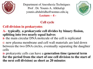
Types of Cell Division: Binary Fission, Mitosis & Meiosis
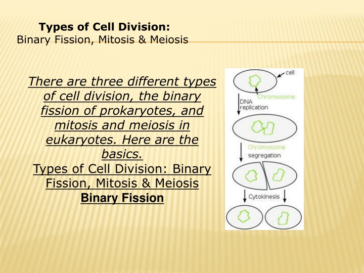
Cell division comes in various forms - binary fission in prokaryotes, and mitosis and meiosis in eukaryotes. While prokaryotes divide through binary fission, eukaryotic cells undergo either mitosis for growth and repair or meiosis for sexual reproduction. Understanding these processes is crucial in the study of genetics and cell biology.

Download Presentation

Please find below an Image/Link to download the presentation.
The content on the website is provided AS IS for your information and personal use only. It may not be sold, licensed, or shared on other websites without obtaining consent from the author.If you encounter any issues during the download, it is possible that the publisher has removed the file from their server.
You are allowed to download the files provided on this website for personal or commercial use, subject to the condition that they are used lawfully. All files are the property of their respective owners.
The content on the website is provided AS IS for your information and personal use only. It may not be sold, licensed, or shared on other websites without obtaining consent from the author.
E N D
Presentation Transcript
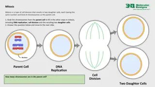
Types of Cell Division: Binary Fission, Mitosis & Meiosis Binary Fission Diagram There are three different types of cell division, the binary fission of prokaryotes, and mitosis and meiosis in eukaryotes. Here are the basics. Types of Cell Division: Binary Fission, Mitosis & Meiosis Binary Fission
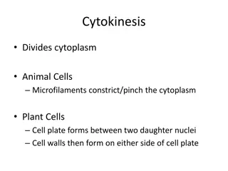
cytokinesis - division of the cytoplasm. This usually occurs witmitosis - nuclear/chemical events resulting in two daughter nuclei which have identical genetic material to each other and to the mother cell h mitosis, but in some organisms this is not so
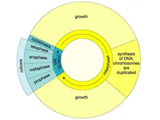
All Organisms Consist of Cells and Arise from Preexisting Cells Mitosis is the process by which new cells are generated. Meiosis is the process by which gametes are generated for reproduction. The Cell Cycle Represents All Phases in the Life of a Cell DNA replication (S phase) must precede mitosis, so that all daughter cells receive the same complement of chromosomes as the parent cell. The gap phases separate mitosis from S phase. This is the time when molecular signals mediate the switch in cellular activity. Mitosis involves the separation of copied chromosomes into separate cells
Chromosome structure Chromosome structure composed of DNA and protein (histones) all tightly wrapped up in one package duplicated chromosomes are connected by a centromere
Example - an organism is 2n = 4. Chromosomes 1 & 2 are homologous chromosomes Chromosomes 3 & 4 are homologous chromosomes Chromosomes 1 & 3 came from the mother Chromosomes 2 & 4 came from the father
Regulation of the Cell Cycle Regulation of the Cell Cycle The cell cycle is controlled by a cyclically operating set of reaction sequences that both trigger and coordinate key events in the cell cycle The cell-cycle control system is driven by a built-in clock that can be adjusted by external stimuli (chemical messages) - a critical control point in the cell cycle where stop and go- ahead signals can regulate the cell cycle Animal cells have built-in stop signals that halt the cell cycles and checkpoints until overridden by go-ahead signals. Three Major checkpoints are found in the G1, G2, and M phases of the cell cycle
The G1 - the Restriction Point The G1 checkpoint ensures that the cell is large enough to divide, and that enough nutrients are available to support the resulting daughter cells. If a cell receives a go-ahead signal at the G1 checkpoint, it will usually continue with the cell cycle If the cell does not receive the go-ahead signal, it will exit the cell cycle and switch to a non-dividing state called G0 Actually, most cells in the human body are in the G0 phase The G2 checkpoint ensures that DNA replication in S phase has been completed successfully. The metaphase checkpoint ensures that all of the chromosomes are attached to the mitotic spindle by a kinetochore
Meiosis Meiosis Meiosis Is a Special Type of Cell Division That Occurs in Sexually Reproducing Organisms Meiosis reduces the chromosome number by half, enabling sexual recombination to occur. Meiosis of diploid cells produces haploid daughter cells, which may function as gametes. Gametes undergo fertilization, restoring the diploid number of chromosomes in the zygote
Meiosis and fertilization introduce genetic variation in three ways: Crossing over between homologous chromosomes at prophase I. Independent assortment of homologous pairs at metaphase I: Each homologous pair can orient in either of two ways at the plane of cell division. The total number of possible outcomes = 2n (n = number of haploid chromosomes). Random chance fertilization between any one female gamete with any other male gamete. The Role of Sexual Reproduction in Evolution
Sexual reproduction in a population should decline in frequency relative to asexual reproduction. Asexual reproduction No males are needed, all individuals can produce offspring. Sexual reproduction Only females can produce offspring, therefore fewer are produced. Sexual reproduction may exist because it provides genetic variability that reduces susceptibility of a population to pathogen attack.
The stages of meiosis can be broken down into two main stages, Meiosis I Meiosis I and Meiosis II Meiosis I Meiosis I can be broken down into four substages: Prophase I, Metaphase I, Anaphase I and Telophase I Meiosis II Meiosis II can be broken down into four substages: Prophase II, Metaphase II, Anaphase II and Telophase II Meiosis II
Meiosis I Meiosis I Prophase I Prophase I - most of the significant processes of Meiosis occur during Prophase I The chromosomes condense and become visible The centrioles form and move toward the poles The nuclear membrane begins to dissolve The homologs pair up, forming a tetrad Each tetrad is comprised of four chromotids - the two homologs, each with their sister chromatid Homologous chromosomes will swap genetic material in a process known as crossing over crossing over (abbreviated as XO) Crossing over serves to increase genetic diversity increase genetic diversity by creating four unique chromatids Compare Prophase I to Prophase II and to the Prophase stage of mitosis.
Metaphase I Metaphase I Microtubules grow from the centrioles and attach to the centromeres The tetrads line up along the cell equator Compare Metaphase I to Metaphase II and to the Metaphase stage of mitosis.
Anaphase I Anaphase I The centromeres break and homologous chromosomes chromosomes separate (note that the sister chromatids chromatids are still attached) Cytokinesis begins Compare Anaphase I to Anaphase II and to the Anaphase stage of mitosis homologous sister
Meiosis II Meiosis II Prophase II Prophase II Centrioles form and move toward the poles The nuclear membrane dissolves Compare Prophase II to Prophase I and to the Prophase stage of mitosis.
Metaphase II Metaphase II Microtubules grow from the centrioles and attach to the centromeres The sister chromatids line up along the cell equator Compare Metaphase II to Metaphase I and to the Metaphase stage of mitosis.
Anaphase II Anaphase II The centromeres break and sister chromatids chromatids separate Cytokinesis begins Compare Anaphase II to Anaphase I and to the Anaphase stage of mitosis. sister
Telophase Telophase II II The chromosomes may decondense (depends on species) Cytokinesis reaches completion, creating four haploid daughter four haploid daughter cells cells Compare Telophase II to Telophase I and to the Telophase stage of mitosis.
Cell Division, Mitosis, and Meiosis Cell Division, Mitosis, and Meiosis Cell Division Functions in Reproduction, Cell Division Functions in Reproduction, Growth, and Repair Growth, and Repair Cell division involves the distribution of identical genetic material, DNA, to two daughters cells. What is most remarkable is the fidelity with which the DNA is passed along, without dilution or error, from one generation to the next.
Lecture 3 Biology Dr.Widad.J.H. Human Body Tissue If you were to try to explain to someone what your body is made of, you might say two arms, two legs, feet and hands, a head and a torso. Or, you might go to the other extreme and say that you are made up of billions of cells. Both answers would be correct. However, there is a more specific way to describe what makes up a body. We are composed of several different types of human body tissue. But what exactly does that mean? The Types of Tissue We have determined that we are made up of four different types of tissue. In addition to muscle tissue, we have connective, epithelial and nervous tissue in the body. So, how are these tissue types different? Let's zoom in on each one to better understand.
Muscle Tissue As mentioned earlier, these different types of tissue are made of particular kinds of cells that work together. First let's look at muscle tissue. Muscle tissue is made up of excitable cells that are long and fibrous. These cells are ready for contraction, or the activation of tension in our muscles, making it possible for us to move our body parts. They are arranged in parallel lines and are bundled, making muscle tissue very strong. If you take a pile of rubber bands, line them up next to each other and attempt to stretch them, you may get the idea of the nature of the muscle tissue.
Cell Structure of Muscle Tissue Cell Structure of Muscle Tissue
Epithelial Tissue Epithelial tissue is made up of epithelial cells, which are vastly different from the muscle cells we just talked about. These cells can be flat, cuboidal, or columnar. They are joined tightly together, making a single or stacked continuous sheet. Like a quilt that is tightly stitched, epithelium makes an excellent protective cover for the body, in the form of skin. Epithelial tissue can also be found lining some internal cavities and organs. Various Configurations of Epithelial Tissue Various Configurations of Epithelial Tissue
Connective Tissue As its name suggests, connective tissue makes up a connective web inside our body. Holding our body parts together and providing support are the main jobs of this tissue. We would certainly not be in good shape if all of our internal body parts were free-floating. Connective tissue fills in the spaces inside our body with a matrix made of fibers within a liquid, solid, or jelly-like substance. Think of a gelatin salad with fruit suspended inside, and you will have an idea of how certain types of connective tissue function.
Nervous Tissue Nervous tissue is found within the nervous system and is made up of unique specialized cells. Like electrical circuits, the nervous system transmits signals from nerves to the spinal cord and brain. Cells known as neurons conduct these impulses, making it possible for us to use our senses. Neuron with Supporting Tissue
Examples of Body Tissue You already know many examples of tissue found in the body. Muscle tissue is found in all of the obvious places, as in our biceps, triceps, quadriceps and so on. But muscle is also located in many of our internal organs, such as the walls of our arteries and digestive tract. And don't forget about the most important muscle of all: the heart. Cardiac tissue is also muscle tissue, as this powerful organ is constantly contracting to pump blood throughout our body.
http://upload.wikimedia.org/wikipedia/commons/thumb/6/64/423_Table_04_02_Summary_of_Epithelial_Tissue_CellsN.jpg/220px-423_Table_04_02_Summary_of_Epithelial_Tissue_CellsN.jpghttp://upload.wikimedia.org/wikipedia/commons/thumb/6/64/423_Table_04_02_Summary_of_Epithelial_Tissue_CellsN.jpg/220px-423_Table_04_02_Summary_of_Epithelial_Tissue_CellsN.jpg Summary showing different epithelial cells/tissues and their characteristics
In general, tissues are classified by the morphology of their cells, and the number of layers they are composed of. Epithelial tissue that is only one cell thick is known as simple epithelium. If it is two or more cells thick, it is known as stratified epithelium However, when taller simple epithelial cells (see columnar, below) are viewed in cross section with several nuclei appearing at different heights, they can be confused with stratified epithelia. This kind of epithelium is therefore described as "pseudostratified" epithelium.
There are three principal morphologies associated with epithelial cells: Squamous epithelium has cells that are wider than they are tall (flat and scale-like). Cuboidal epithelium has cells whose height and width are approximately the same (cube shaped). Columnar epithelium has cells taller than they are wide (column-shaped). In addition, the morphology of the cells in transitional epithelium may vary from squamous to cuboidal, depending on the amount of tension on the epithelium. Simple epithelium Simple epithelium Simple epithelium is one cell thick; that is, every cell is in direct contact with the underlying basement membrane. In general, it is found where absorption and filtration occur. The thinness of the epithelial barrier facilitates these processes.
(1) simple squamous; which is found lining areas where passive diffusion of gases occur. e.g. walls of capillaries, linings of the pericardial, pleural,and peritoneal cavities, as well as the linings of the alveoli of the lungs. (2) simple cuboidal: these cells may have secretory, absorptive, or excretory functions. examples include small collecting ducts of kidney,pancreas and salivary gland. (3) simple columnar; found in areas with extremely high secretive (as in wall of the stomach), or absorptive (as in small intestine) areas. they possess cellular extensions (e.g. microvilli in the small intestine, or cilia found almost exclusively in the female reproductive tract). (4) pseudostratified epithelia; they are also called respiratory epithelium. this is due to their almost exclusive confinement to the larger respiratory airways i.e. the nasal cavity, trachea, bronchi e.t.c
Stratified epithelium Stratified epithelium Stratified epithelium differs from simple epithelium in that it is multilayered. It is therefore found where body linings have to withstand mechanical or chemical insult such that layers can be abraded and lost without exposing subepithelial layers. Cells flatten as the layers become more apical, though in their most basal layers the cells can be squamous, cuboidal or columnar. Stratified epithelial tissue also differs from simple epithelial tissue in that stratified epithelial tissues do not contain junctional complexes, and have their cells bound together only by desmosomes. Stratified epithelia (of columnar, cuboidal or squamous type) can have the following specializations:
Specialization Description In this particular case, the most apical layers (exterior) of cells are dead and lose their nucleus and cytoplasm, instead contain a tough, resistant protein called keratin. This specialization makes the epithelium waterproof, so is found in the mammalian skin. The lining of the esophagus is an example of a non-keratinized or "moist" stratified epithelium.[10] Transitional epithelia are found in tissues that stretch and it can appear to be stratified cuboidal when the tissue is not stretched or stratified squamous when the organ is distended and the tissue stretches. It is sometimes called the urothelium since it is almost exclusively found in the bladder, ureters and urethra.[10] Keratinized Transitional
Structure Structure Cells of epithelial tissue are tightly packed and form a continuous sheet. They have almost no intercellular spaces. All epithelia is usually separated from underlying tissues by an extra cellular fibrous basement membrane. The lining of the mouth, lung alveoli and kidney tubules all are made of epithelial tissue. The lining of the blood and lymphatic vessels are of a specialised form of epithelium called endothelium
Basement membrane Basement membrane Epithelial tissue rests on a basement membrane, which acts as a scaffolding on which epithelium can grow and regenerate after injuries.[11]Epithelial tissue is innervated, but avascular. This epithelial tissue must be nourished by substances diffusing from the blood vessels in the underlying tissue, but they don't have their own blood supply. The basement membrane acts as a selectively permeable membrane that determines which substances will be able to enter the epithelium. Cell junctions Cell junctions Cell junctions are especially abundant in epithelial tissues. They consist of protein complexes and provide contact between neighbouring cells, between a cell and the extracellular matrix, or they build up the paracellular barrier of epithelia and control the paracellular transport
Cell junctions are the contact points between plasma membrane and tissue cells. There are mainly 5 different types of cell junctions: tight junctions, adherens junctions, desmosomes, hemidesmosomes, and gap junctions. Tight junctions are a pair of trans-membrane protein fused on outer plasma membrane. Adherens junctions are a plaque (protein layer on the inside plasma membrane) which attaches both cells' microfilaments. Desmosomes attach to the microfilaments of cytoskeleton made up of keratin protein. Hemidesmosomes resemble desmosomes on a section. They are made up of the integrin (a transmembrane protein) instead of cadherin. They attach the epithelial cell to the basement membrane. Gap junctions connect the cytoplasm of two cells and are made up of proteins called connexins (six of which come together to make a connexon).
Development Development Epithelial tissues are derived from all of the embryological germ layers:[ from ectoderm (e.g., the epidermis); from endoderm (e.g., the lining of the gastrointestinal tract); from mesoderm (e.g., the inner linings of body cavities). However, it is important to note that pathologists do not consider endothelium and mesothelium (both derived from mesoderm) to be true epithelium. This is because such tissues present very different pathology. For that reason, pathologists label cancers in endothelium and mesothelium sarcomas, whereas true epithelial cancers are called carcinomas. Also, the filaments that support these mesoderm- derived tissues are very distinct. Outside of the field of pathology, it is, in general, accepted that the epithelium arises from all three germ layers
Secretory Secretory epithelia epithelia Different forms of secretion is possible in epithelial tissue or in glands. http://upload.wikimedia.org/wikipedia/commons/thumb/3/37/405_Modes_of_Secretion_by_Glands_updated.svg/220px-405_Modes_of_Secretion_by_Glands_updated.svg.png
Glands are formed from the invagination/infolding of epithelium and subsequent growth in the underlying connective tissue. There are two major classifications of glands: endocrine glands and exocrine glands. Endocrine glands secrete their product into the extracellular space where it is rapidly taken up by the blood vascular system. Exocrine glands secrete their products into a duct that then delivers the product to the lumen of an organ or onto the free surface of the epithelium. In arthropods, the integument, or external "skin", consists of a single layer of epithelial ectoderm from which arises the cuticle,[13]an outer covering of chitin the rigidity of which varies as per its chemical composition.
http://upload.wikimedia.org/wikipedia/commons/thumb/5/5b/406_Types_of_Glands.jpg/220px-406_Types_of_Glands.jpghttp://upload.wikimedia.org/wikipedia/commons/thumb/5/5b/406_Types_of_Glands.jpg/220px-406_Types_of_Glands.jpg Different characteristics of glands of the body
Glands are formed from the invagination/infolding of epithelium and subsequent growth in the underlying connective tissue. There are two major classifications of glands: endocrine glands and exocrine glands. Endocrine glands secrete their product into the extracellular space where it is rapidly taken up by the blood vascular system. Exocrine glands secrete their products into a duct that then delivers the product to the lumen of an organ or onto the free surface of the epithelium. In arthropods, the integument, or external "skin", consists of a single layer of epithelial ectoderm from which arises the cuticle, an outer covering of chitin the rigidity of which varies as per its chemical composition.
Sensing the extracellular environment Sensing the extracellular environment "Some epithelial cells are ciliated, and they commonly exist as a sheet of polarised cells forming a tube or tubule with cilia projecting into the lumen." Primary cilia on epithelial cells provide chemosensation, thermosensation, and mechanosensation of the extracellular environment by playing "a sensory role mediating specific signalling cues, including soluble factors in the external cell environment, a secretory role in which a soluble protein is released to have an effect downstream of the fluid flow, and mediation of fluid flow if the cilia are motile


 simple squamous; which is found lining areas where")