Types of Electron Microscopes and Microscopy Techniques
Explore the differences between Scanning Electron Microscope (SEM) and Transmission Electron Microscope (TEM). Learn about new microscopy techniques like Confocal Microscopy and the comparison between Light Microscope and Electron Microscope. Discover how SEM and TEM work and their applications in studying microscopic structures.

Download Presentation

Please find below an Image/Link to download the presentation.
The content on the website is provided AS IS for your information and personal use only. It may not be sold, licensed, or shared on other websites without obtaining consent from the author. If you encounter any issues during the download, it is possible that the publisher has removed the file from their server.
You are allowed to download the files provided on this website for personal or commercial use, subject to the condition that they are used lawfully. All files are the property of their respective owners.
The content on the website is provided AS IS for your information and personal use only. It may not be sold, licensed, or shared on other websites without obtaining consent from the author.
E N D
Presentation Transcript
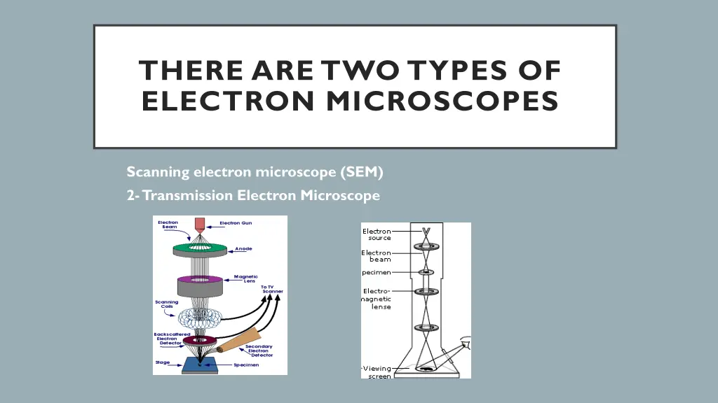
THERE ARE TWO TYPES OF ELECTRON MICROSCOPES 1. 2. Scanning electron microscope (SEM) 2-Transmission Electron Microscope
NEW TECHNIQUES IN MICROSCOPY: Confocal Microscopy (Confocal Scanning Laser Microscope)
Light Microscope Electron Microscope Cheap to purchase Expensive to buy Cheap to operate. Expensive to produce electron beam. Small and portable. Large and requires special rooms. Simple and easy sample preparation. Lengthy and complex sample prep. Material rarely distorted by preparation. Preparation distorts material. Vacuum is not required. Vacuum is required. Natural color of sample maintained. All images in black and white.
Transmission Electron Microscope (TEM) Pass a beam of electrons through the specimen. The electrons that pass through the specimen are detected on a fluorescent screen on which the image is displayed. Thin sections of specimen are needed for transmission electron microscopy as the electrons have to pass through the specimen for the image to be produced. This is the most common form of electron microscope and has the best resolution Bacterium (TEM)
Scanning Electron Microscope (SEM) Pass a beam of electrons over the surface of the specimen in the form of a scanning beam. Electrons are reflected off the surface of the specimen as it has been previously coated in heavy metals. It is these reflected electron beams that are focused of the fluorescent screen in order to make up the image. Larger, thicker structures can thus be seen under the SEM as the electrons do not have to pass through the sample in order to form the image. This gives excellent 3-dimensional images of surfaces However the resolution of the SEM is lower than that of the TEM. A head and the right eye of a fly (SEM)


")
")