Ultrasound Imaging: An Overview of Uses and Benefits
Ultrasound imaging, also known as sonography, utilizes high-frequency sound waves to visualize internal organs without radiation. It is a widely used and cost-effective imaging technology applied in various medical fields including obstetrics, cardiology, and urology. This non-invasive procedure can assist in diagnosing a range of conditions and evaluating organ damage, making it a valuable tool in modern healthcare.

Uploaded on Apr 19, 2025 | 1 Views
Download Presentation

Please find below an Image/Link to download the presentation.
The content on the website is provided AS IS for your information and personal use only. It may not be sold, licensed, or shared on other websites without obtaining consent from the author.If you encounter any issues during the download, it is possible that the publisher has removed the file from their server.
You are allowed to download the files provided on this website for personal or commercial use, subject to the condition that they are used lawfully. All files are the property of their respective owners.
The content on the website is provided AS IS for your information and personal use only. It may not be sold, licensed, or shared on other websites without obtaining consent from the author.
E N D
Presentation Transcript
www.studymafia.org Seminar On Ultrasound Submitted To: www.studymafia.org www.studymafia.org Submitted By:
Outline What is Ultrasoundimaging? WhyUltrasound? CommonUses History Properties ofUltrasound Equipment How does the procedurework? Benefits andRisks
What is General UltrasoundImaging? Ultrasound imaging, also called sonography, involves exposing part of the body to high- frequency sound waves to produce pictures ofthe inside of thebody. Ultrasound examinations do not useionizing radiation (as used in x-rays). Because ultrasound images are captured in real- time, they can show the structure and movementof the body's internal organs, as well as blood flowing through bloodvessels.
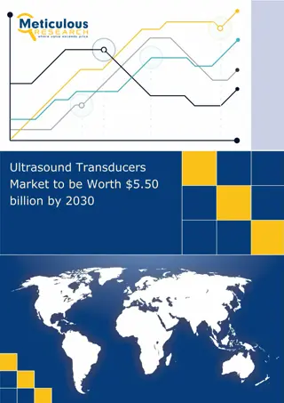
WhyUltrasound Ultrasound (US) is the most widelyused imaging technologyworldwide Popular due to availability, speed, lowcost, patient-friendliness (noradiation) Applied in obstetrics, cardiology,inner medicine,urology,... Ongoing research to improve image quality, speed and new application areas such a intra- operative navigation, tumourtherapy,...
Whataresomecommonusesoftheprocedure? 1.Ultrasound examinations can help to diagnose a variety of conditions and to assess organ damage following illness. 2.Ultrasound is used to help physiciansevaluate symptoms suchas: pain swelling infection hematuria (blood inurine)
Ultrasound is a useful way of examining many of thebody's internal organs,including but not limited to the: heart and blood vessels, including the abdominal aortaand its majorbranches Liver Gallbladder Spleen Pancreas Kidneys Bladder Uterus, ovaries, and unborn child (fetus) inpregnant patients Eyes Thyroid and parathyroidglands Scrotum(testicles) brain ininfants hips in infants
Ultrasound is also usedto: guide procedures such asneedle biopsies, in which needles are used to extract samplecells from an abnormal area for laboratorytesting. image the breasts and to guidebiopsyofbreast cancer diagnose a variety of heart conditions and to assess damage after a heart attack or diagnose for valvular heartdisease.
Doppler ultrasound images can help thephysician to see andevaluate: blockages to blood flow (such asclots). narrowing of vessels (which may be causedby plaque). tumors and congenital vascularmalformation. With knowledge about the speed and volumeof blood flow gained from a Doppler ultrasound image, the physician can often determine whether a patient is a good candidate for a procedure likeangioplasty.
Applications of ultrasound: Few applications of ultrasound are mentioned below and it is used for examining the following things: Extremities like connective tissues, muscles, joints, and vessels Neck: vessels, abscesses, thyroid gland, and lymph nodes Chest: heart (echocardiography), wall, pleura, mediastinal tumors, and disorders of lungs which are situated in peripheral area of lungs Abdomen, small pelvis, and retroperitoneum: fluid-containing structures, parenchymatous organs, gastrointestinal tract, great vessels, lymph nodes, abnormal fluid collections, and tumors Spine in infants Hips in infants Brain in infants Eyes Unborn child fetus in pregnant women
History 1877: Lord Raleigh - "Theory ofSound" 1880: Pierre & Jacques Curie -Piezoelectric effect 1914: Langevin - First Ultrasoundgenerator using piezoelectriceffect 1928: Solokov - Ultrasound for materialtesting
1942: Dussik - First application ofUltrasound in medicaldiagnostics Shortly after WWII, researchers inJapan began to explore medical diagnostic capabilities of ultrasound. ... different medical applications (gallstones, tumours) End of 1960's: Boom of Ultrasound inmedical diagnostics
Pan-Scanner -The transducer rotated in a semicirculararc around the patient(1957)
Scan converter allowed for the first time to use the upcoming computer technology to improveUS
Early 1970s Gray scale static images of internalorgans Mid 1970s Real-timeimaging Early 1980s SpectralDoppler ColorDoppler Also produced was a hand-held contact scanner for clinicaluse.
Properties ofUltrasound The frequencies of medical Ultrasound waves are several magnitudes higher than the upper limit of human hearing. Approximate frequency ranges of sound
Although ultrasound is better known for its diagnostic capabilities, it was initially usedfor therapy rather thandiagnosis. In the 1940s, ultrasound was used to perform services similar to that of radiation or chemotherapynow. Ultrasonic waves emit heat that can create disruptive effects on animal tissue anddestroy malignanttissue.
Common SoundFrequencies Sound Frequency 15 20 000Hz Up to 40 000Hz 100 1 500Hz 150 2 500Hz 44 0Hz 50 000 200 000Hz 2.5 40MHz 600MHz Adult audiblerange Range for children'shearing Male speakingvoice Female speakingvoice Standard pitch (ConcertA) Bat MedicalUltrasound Maximum soundfrequency Common sound frequencies and frequency ranges
Physics of themethod Longitudinal mechanicalwaves Needs elasticmedium Transducerneedsto bein contactwith skin Componentresolution 3 MHZ ->1.1 mm 10 MHZ -> .3 mm Wavevelocity Fat -> 1450 m/s Muscle ->1580m/s
Principles ofUltrasound ItsComponents Operations Applications
The Ultrasound Machine A basic ultrasound machine has the followingparts: 1. Transducer probe - probe that sends and receives thesound waves 2. Central processing unit (CPU) - computer that does all of the calculations and contains the electrical power supplies for itself and the transducer probe 3. Transducer pulse controls - changes the amplitude, frequency and duration of the pulses emitted from the transducerprobe 4. Display - displays the image from the ultrasound data processed by the CPU 5. Keyboard/cursor - inputs data and takes measurementsfrom the display 6. Disk storage device (hard, floppy, CD) - stores the acquired images 7. Printer - prints the image from the displayeddata
Equipment Ultrasound scanners consist of a console containing a computer and electronics, a video display screen and atransducerthat is used todo the scanning. The transducer is a small hand-held device that resembles a microphone, attached to thescanner by acord. The transducer sends out inaudible high frequency sound waves into the body and then listensfor the returning echoesfrom the tissues in thebody. The principles are similar to sonar used byboats andsubmarines.
The ultrasound image is immediately visible on a video display screen that looks like a computer or televisionmonitor. The image is created based on the amplitude (strength), frequency and time ittakes for the sound signal to return from the area of the patient being examined to the transducerand the type of body structure the sound travels through.
What are the benefits vs.risks? Benefits Most ultrasound scanning is noninvasive (no needlesor injections) and is usuallypainless. Ultrasound is widely available, easy-to-use and less expensive than other imagingmethods. Ultrasound imaging does not use any ionizingradiation. Ultrasound scanning gives a clear picture of soft tissuesthat do not show up well onx-ray images. Ultrasound is the preferred imagingmodality for the diagnosis and monitoring of pregnant women andtheir unbornbabies. Ultrasound provides real-time imaging, making it a goodtool for guidingminimally invasiveprocedures such asneedle biopsies andneedleaspiration.
Risks For standard diagnostic ultrasound there are no known harmful effects on humans. Unlike X-rays, ultrasound involves onlysound waves No radiationdanger However, sound waves can increase body temperature This is known ascavitation Significant only for long exposuretime
Many studies have been conducted to determinethe physiological effects of ultrasound cavitation No direct correlation has been found between ultrasound imaging and cancer, low birth weight, dyslexia or delayedspeech development Reliable data from ultrasound techniquesis hard to comeby Additional studiesare ongoing Biggest risk ismisdiagnosis
Future ofUltrasound Improved clarity for usein cancerdiagnosis Increased therapeutic use to correct blood clots and kidneystones Portability andveterinary use Joint and muscletreatment through cavitation
What are the limitations of General UltrasoundImaging? Ultrasound waves are disrupted by air or gas; therefore ultrasound is notan ideal imaging technique for air-filled bowel or organs obscured by the bowel. In most cases, barium exams, CT scanning, and MRI are the methods of choice in thissetting. Large patients are more difficult to image by ultrasound because greater amounts of tissue attenuates (weakens) the soundwavesasthey passdeeper into thebody.
Reference www.google.com www.wikipedia.org www.studymafia.com