Understanding Acute Flaccid Paralysis and Its Clinical Manifestations
Explore the definition, epidemiology, pattern of weakness, neuroanatomical differentials, etiology, and clinical clues of Acute Flaccid Paralysis (AFP) syndrome, as defined by the World Health Organization (WHO). Learn about the rapid onset of weakness, absence of spasticity, and various causes of AFP, such as viral infections, toxins, trauma, and neuromuscular disorders.

Download Presentation

Please find below an Image/Link to download the presentation.
The content on the website is provided AS IS for your information and personal use only. It may not be sold, licensed, or shared on other websites without obtaining consent from the author. If you encounter any issues during the download, it is possible that the publisher has removed the file from their server.
You are allowed to download the files provided on this website for personal or commercial use, subject to the condition that they are used lawfully. All files are the property of their respective owners.
The content on the website is provided AS IS for your information and personal use only. It may not be sold, licensed, or shared on other websites without obtaining consent from the author.
E N D
Presentation Transcript
ACUTE FLACCID PARALYSIS BY MBBSPPT.COM (AFP)
DEFINITION (WHO) WHO defines AFP syndrome as characterized by rapid onset of weakness of an individual s extremities, often including weakness of the muscles of respiration and swallowing, progressing to maximum severity within 1-10 days. The term flaccid indicates the absence of spasticity or other signs of disordered central nervous system (CNS) motor tracts such as hyperreflexia, clonus, or extensor plantar responses (World Health Organization 1993)
EPIDEMIOLOGICAL DEFINITION (WHO 2005) Clinical case definition Any child under 15 years of age with AFP (due to any cause) or any person of any age with paralytic illness if polio is suspected
PATTERN OF WEAKNESS Sign Umn Lmn Myopathic Atrophy - +++ + Fasciculations - - +++ Tone +++ - +/- Distribution Regional Segmental Proximal Tendon Reflexes -/-- +/- +++ Babinski s Sign - - +
NEUROANATOMICAL D/D OF AFP Spinal Cord Compression: Traumatic, Epidural Abscess Inflammatory: Transverse Myelitis Ant Horn Cell Viral: Polio, Enetovirus, JE Vascular: Ant Spinal Artery Occlusion Roots/Nerves: Immune Mediated: GBS
CONTINUED Toxin: Post Diphtheria, Porphyria, Arsenic, Heavy Metals Viral: Rabies Trauma: I/M Injection Neuromuscular Junction Immune Mediated: Mysthenia Gravis Drugs And Toxins: Organophosphorus, Snake Venom, Aminoglycoside, Botulinium, Tick
CONTINUED Muscle Infection: Viral Myositis Inflammation: Polymyositis Channelopathy: Hypokalemia Dyselectronemia: Hypokalemia
CLINICAL CLUES (AFP) Fever At Onset: Polio or Enteroviral Myelitis, TM, Myositis, Potts Spine Trauma: Compression of Spinal Cord Exposure: Lead, Arsenic, Tick, Snake Venoms Preceding Infection: GBS, TM Precipitating Factors: Diarrhea (Hypokalemia) Intramuscular Injection: Traumatic Neuritis, Polio
CLINICAL CLUES (AFP) Sensory level/bladder bowel: compressive myelopathy, TM Constipation < 1 yr: botulinism (honey injestion) Ascending weakness: GBS, Rabies, VZV Descending weakness: Diphtheria, botulinism Ptosis: Mysthenia gravis, botulinism DTR: Absent (GBS, TM early stage, polio) Preserved: Myasthenia, periodic paralysis
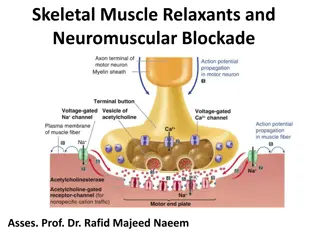
GUILLAIN-BARR SYNDROME Acute idiopathic monophasic acquired inflammatory demyelinating polyradiculoneuropathy. Most common cause of AFP in healthy infants and children. An acute, rapidly progressing and potentially fatal form of polyneuritis
PATHOPHYSIOLOGY Autoimmune disorder (T cell sensitization) Cause of demyelization Due to attack of the myelin sheath of nerves by: Antibodies (Ig M, Ig G) White Blood Cells (Macrophages) Complement activation Virus/Bacteria share antigenic sites with axons & peripheral nerve sheath or both
PATHOPHYSIOLOGY (CONTINUED) Leakage of proteins into the CSF causing raised CSF proteins without pleocytosis Can involve the peripheral nerves, cranial nerves, dorsal roots, dorsal root ganglia & sympathetic chain
AETIOLOGY Bacteria Mycoplasma Compylobacter Jejuni Virus Hepatitis B HSV EBV Measles Mumps Echovirus Cocksakie virus Influenza virus Varicella virus
CLASSIFICATION OF GBS AIDP: most common form Classic type (mixed) Pure motor axonal GBS: Strong C.jejuni association, common in India Pure sensory axonal GBS Pure pandysautonomia Miller-Fisher syndrome( hypotonia, ophthalmoplegia, ataxia)
GUILLAIN-BARRE SYNDROME Affects the peripheral nervous system
CLINICAL FEATURES Motor: 1- Symmetric acute progressive ascending weakness <4 wks, starting in LL 2- Areflexia or hyporeflexia 3- Atonia or hypotonia Sensation: 1- C/O pain as hyperthesia orcramps 2- O/E loss of pain sensation (hypothesia) in feet/hands Autonomic: orthostatic hypotension, cardiac arrythmia
CHARACTERISTIC 3ATRIAD: Ascending Weakness: Bilateral Symmetrical Weakness, LL>UL Others - Cranial Nerves (Brain Stem) - Respiratory Muscles - Phrenic Nerves( Diaphragm ) Areflexia( Hallmark) Atonia( Hypotonia)
SERIOUS ASSOCIATION: Respiratory Failure: Diaphragmatic Weakness (Phrenic Nerves.) Respiratory Muscles Weakness Oropharyngeal Weakness: Impaired Swallowing Of Secretions & Aspiration Cardiac Arrest Aspiration Pneumonia
INVESTIGATIONS Early Nerve Conduction Velocity (NCV) abnormality: most specific test AFTER 1ST WEEK Late: CSF study: albuminocytogenic dissociation Others Antibody study: Ig M autoantibodies to GM1 and GM2 gangliosides Anti-GQ1b antibodies
DOUBT THE DIAGNOSIS OF GBS IF Marked persistent asymmetry of weakness. Persistent bladder or bowel dysfunction. Bladder or bowel dysfunction at the onset. Mononuclear leukocytosis in the CSF > 50. Sharp sensory level.
TREATMENT Hospitalization General care Specific treatment Treatment of complication
GBS MANAGEMENT Critical care monitoring autonomic and respiratory dysfunction Admission criteria in PICU: A. Flaccid Quadriparesis B. Rapidly Progressive Weakness C. Reduced Vital Capacity ( 20 Ml/Kg) D. Bulbar Palsy E. Autonomic Cardiovascular Instability
SPECIAL THERAPY Immune modulatory therapy: Intravenous Immunoglobulins Plasmapheresis Both therapies have been shown to shorten recovery time by as much 50%.
GBS MANAGEMENT Risk factors for respiratory failure in GBS: Cranial nerve involvement. Short time from preceding respiratory illness. Rapid progression over less than 7 days. Elevated CSF protein in the first week. Severe weakness 20% of children with GBS require mechanical ventilation for respiratory failure.
OUTCOME OF GBS PATIENTS Regressive: 90 % of patient's good prognosis (descending pattern recovery) Chronic Relapsing: Less than 5% of patients Mortality: 3% die from complications as respiratory failure especially in infants
TRANSVERSE MYELITIS (DIAGNOSTIC CRITERIA) Sensory, motor, or autonomic dysfunction attributable to the spinal cord Bilateral signs and/or symptoms Clearly defined sensory level Inflammation defined by CSF pleocytosis or elevated IgG index or gadolinium enhancement MRI Progression to nadir between 4 hours and 21 days.
PATHOPHYSIOLOGY Idiopathic (75-90%) Vs Disease Associated TM Abnormal Activation of The Immune System Resulting In Inflammation and Injury Within The Spinal Cord. Para infectious Etiology Antigenic Mimicry
CLINICAL FEATURES Mean age of onset is 9 years. Symptoms progress rapidly, peaking within 2 days. Usually, level of myelitis is thoracic. Symmetrical leg weakness, sensory level and early bladder involvement. Recovery usually begins after a week of onset.
PHYSICAL EXAMINATION Tenderness over the spine may point to trauma or infection. Visual acuity and color vision, funduscopic examination for optic neuritis. Increased tone, spastic weakness, LL>UL Reflexes are usually brisk, with positive Babinski sign. Sensory ataxia, a sensory level. Sphincter dysfunction.
DIAGNOSIS MRI and CSF analysis are the two most important tests and are mandatory. If both tests are negative, repeat tests in 2 to 7 days is recommended. MRI of spine: usually shows swelling of the cord, but at times is normal.
LUMBAR PUNCTURE Normal or slightly increased protein. Mild pleocytosis with lymphocyte predominance. Elevation of IgG index and presence of oligoclonalbands are indicative of MS or other systemic inflammatory disease. CSF for infections: all should be negative. Investigation for underlying systemic inflammatory disorder.
TREATMENT IV methylprednisolone (DOC) IV immunoglobulin (IVIG) or plasmapheresis Cyclophosphamide has been reported to be useful in myelitis associated with systemic inflammatory diseases.
TREATMENTCONT Symptomatic management of bowel and bladder dysfunction. Management of respiratory, cardiovascular & autonomic dysfunction. Physical and occupational therapy (PT/OT) may help promote functional recovery and prevent contractures.
PROGNOSIS Variable prognosis ,depends upon the severity of involvement 33% recovery complete, 33% moderate disability, 33% severe disability Recovery can take up to 2 yr, mostly within 3-6 months
Clinical feature GBS TM Onset of paralysis Motor findings Hours to 4 weeks Ascending weakness Within 4 days Para paresis or quadriparesis Spinal cord levels Sensory findings Ascending sensory Autonomous findings CVS Bladder and bowel Cranial nerves EOM palsies or facial weakness None CSF Pleocytosis uncommon, raised protein Pleocytosis Common, protein normal Focal abnormalities MRI Normal NCV Diagnostic test MRI spine
MANAGEMENT PROTOCOLS OF AFP Step 1: ABC & electrolytes Step 2: examination and classification - AFP with sensory level: TM, compressive myelopathy - Afebrile symmetric AFP: (+/- bulbar & respiratory involvement) + areflexia& minimal sensory loss( sensory symptoms present): GBS
MANAGEMENT PROTOCOLS Febrile pure motor asymmetric AFP( no bladder involvement) with meningismus: polio & nonpolio enetrovirus Flaccid motor-sensory LL monoparesis after IM injection: injection neuritis Opthalmoplegia, descending paralysis, ataxia, areflexia: millar fisher variant GBS Proximal muscle weakness with normal DTR: Viral myositis, inflammatory myopathy
MANAGEMENT( CONTINUED) Investigation: MRI: all cases of compressive myelopathy, TM Xray Spine: suspected atlanto-axial dislocation, pott s spine NCV Testing: GBS LP: GBS, TM,Viral myelitis Biochemistry: CK, K,Ca, Po4 Urine for porphyria, arsenic, lead
MANAGEMENT( CONTINUED) General: Meticulous supportive care, anticipate & management of respiratory & bulbar weakness, shock, autonomic instability, prevention of nosocomial infections & bed sores Specific therapy: IVIG: GBS IV methyl prednisolone: TM Hypokalemia: IV k correction
SUMMARY AFP is a broad clinical entity Every case of AFP has to be reported Every case is a medical emergency Accurate & early diagnosis of the cause is important Early detection & management of complications are critical

")


")
")


")


")
")