Understanding Autonomic Nervous System Pathway
In the autonomic nervous system pathway, two neurons synapse in an autonomic ganglion. Pre- and postganglionic neurons have specific characteristics and are part of the sympathetic and parasympathetic divisions. Reflex arcs in the nervous system demonstrate automatic responses to sensory stimuli, involving both sensory and motor neurons. Sensory nerve endings play a crucial role in perceiving stimuli and generating nerve impulses. Different modalities of sensation are perceived and classified by the brain.

Download Presentation

Please find below an Image/Link to download the presentation.
The content on the website is provided AS IS for your information and personal use only. It may not be sold, licensed, or shared on other websites without obtaining consent from the author.If you encounter any issues during the download, it is possible that the publisher has removed the file from their server.
You are allowed to download the files provided on this website for personal or commercial use, subject to the condition that they are used lawfully. All files are the property of their respective owners.
The content on the website is provided AS IS for your information and personal use only. It may not be sold, licensed, or shared on other websites without obtaining consent from the author.
E N D
Presentation Transcript
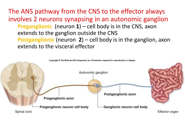
The ANS pathway from the CNS to the effector always involves 2 neurons synapsing in an autonomic ganglion Preganglionic (neuron 1) cell body is in the CNS, axon extends to the ganglion outside the CNS Postganglionic (neuron 2) cell body is in the ganglion, axon extends to the visceral effector
Preganglionic (neuron #1) Always myelinated Neurotransmitter is always ACh Postganglionic (neuron #2) Always non myelinated Neurotransmitter is Ach or norepinephrine Sympathetic division (thoracolumbar) Cell bodies for all the neurons #1 reside in the thoracic and lumbar portions of the spinal cord. Parasympathetic division (craniosacral) Cell bodies reside in the brain stem (cranial nerves) or in the sacral portion of the spinal cord.
The reflex: In general, nerve function is dependent on both sensory and motor fibers, sensory stimulation evoking motor response. Even the autonomic system is activated by sensory impulses from receptors in the organ or muscle. Where especially sensitive areas or powerful stimuli are concerned, it is not always necessary for a sensory impulse to reach the brain in order to trigger motor response. A sensory neuron may link directly to a motor neuron at a synapse in the spinal cord, forming a reflex arc that performs automatically. Thus, tapping the tendon below the kneecap causes the leg to jerk involuntarily because the impulse provoked by the tap, after traveling to the spinal cord, travels directly back to the leg muscle. Such a response is called an involuntary reflex action.
Commonly, the reflex arc includes one or more connector neurons that exert a modulating effect, allowing varying degrees of response, e.g., according to whether the stimulation is strong, weak, or prolonged. Reflex arcs are often linked with other arcs by nerve fibers in the spinal cord. Consequently, a number of reflex muscle responses may be triggered simultaneously, as when a person shudders and jerks away from the touch of an insect.
Sensory Nerve Endings Sensory nerve endings may be classified functionally or morphologically. Functional classification It is important to distinguish between a stimulus and the sensation it elicits. A nerve ending perceives a stimulus and generates a nerve impulse. It is perceived as a sensation by the brain. The following are different modalities of sensation.
Pain Temperature Touch (crude touch or fine touch) Discriminatory touch Stereognosis Vibration Proprioception
Morphological classification Nerve endings are classified according to their structure. Nerve endings are always in contact with a transducer cell that converts a mechanical, thermal or chemical stimulus to an electrical potential in the nerve ending. A- Free nerve endings are the branched terminations of the axons. Myelinated nerves lose their myelin sheath, and end in a number of branches that penetrate the area being innervated. The axon terminals end in small expansions (Merkel s discs) that are in contact with specialized epithelial cells (Merkel s cells or tactile domes). They are very sensitive to pain, temperature and crude touch. Free nerve endings are also common in somatic tissues including bones, joints and muscle.
B- Encapsulated nerve endings consist of branched axon enclosed in a discrete connective tissue capsule. There are several distinct varieties that are found in specific locations and subserve specific functions. 1-Meissner s corpuscles o Are found in thick skin of the palms and soles, and in the skin of the nipples and genitalia. o Are sensitive to discriminatory touch. o Are critically located in the dermal papillae where the overlying epidermis is thinnest o Are oval structures o Have branched, unmyelinated nerve terminals within their core
2-Pacinian corpuscles o Are found in the deep tissues, particularly in the deep layers of the dermis, subcutaneous tissue, around joints, in the parietal pleura and peritoneum o Are sensitive to deep touch, pressure and vibration o Are large structures measuring 1-2 mm in diameter o Have branched, unmyelinated nerve terminals in the core of the corpuscle 3-Ruffini nerve endings o Are stretch receptors o Are located in the dermis of hairy skin o Have a coreof nerve endings and collagen bundles o Are surrounded by a cellular capsule
Nerve endings on smooth muscle, cardiac muscle and secretory cells. o These are the nerve endings of postganglionic autonomic nerves. o They are always free nerve endings and do not have motor end plates or other specialised endings o They may be cholinergic or adrenergic o Gastrointestinal smooth muscle and cardiac muscle contract independently of motor nerve stimulation. The autonomic endings on these two types of muscle may affect the rate of contraction of the muscles.
Alert behavior : When the nervous system is on high alert or in "stress mode" for prolonged periods of time it can place extra pressure on our adrenal glands. The adrenal glands provide the hormonal charge necessary for the fight flight response. When the adrenals glands are depleted we have less energy and tire easily.

")