Understanding Cesarean Section Procedure
A comprehensive overview of Cesarean section (C-section) procedure, including its definition, historical background, rising incidence, indications, and common scenarios where it is performed. Learn about the absolute and relative indications for C-sections, along with the common medical conditions and obstetric history factors that may lead to this surgical intervention. Stay informed about the evolving trends and reasons for the increasing rates of C-section deliveries in recent years.

Download Presentation

Please find below an Image/Link to download the presentation.
The content on the website is provided AS IS for your information and personal use only. It may not be sold, licensed, or shared on other websites without obtaining consent from the author. If you encounter any issues during the download, it is possible that the publisher has removed the file from their server.
You are allowed to download the files provided on this website for personal or commercial use, subject to the condition that they are used lawfully. All files are the property of their respective owners.
The content on the website is provided AS IS for your information and personal use only. It may not be sold, licensed, or shared on other websites without obtaining consent from the author.
E N D
Presentation Transcript
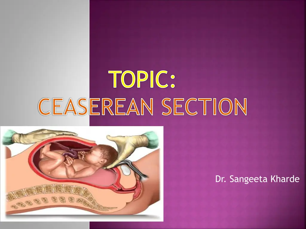
TOPIC: CEASEREAN SECTION Dr. Sangeeta Kharde
DEFINITION: It is an operative procedure whereby the fetuses after the end of 28th wk are delivered through an incision on the abdominal and uterine walls.
HISTORY: Operation derives its name from the notification lex cesarea a Roman law promulgated in 715 B.C. Law states that either an abdominal delivery in a dying woman with a hope to get a live baby or to perform postmortem abdominal delivery. Its derived from Latin verb Caedere which means to cut. French obstetrician, Francois Mauriceau first reported CS in 1668. In 1876, Porro performed subtotal hysterectomy. It was Max Sanger in 1882, first sutured the uterine walls.
Contd., In 1907, Frank described the extraperitoneal operation. Kronig in 1912, introduced lower segment vertical incision & it was popularized by DeLee (1922). In 1881 Kehrer did the transverse lower segment operation for the first time. Munro Kerr in 1926 popularized the present technique of lower segment operation.
INCIDENCE: Its rising. During last decade there has been two to three fold rise in the incidence from the initial rate of about 10%.
INDICATIONS: ABSOLUTE: Severe degree of contracted pelvis with true conjugate < 7.5cm (3 ). Central placenta praevia. Cervical or broad ligament fibroid. Vaginal atresia. Advanced carcinoma cervix.
RELATIVE: Cephalopelvic disproportion and contracted pelvis. Previous uterine scar. Previous scar following classical or hysterotomy. Fetal distress during first stage of labor. Abnormal uterine contractions. APH- placenta praevia, abruptio placentae. Malpresentation.
Contd., Bad obstetric history. Hypertensive disorders. Failed surgical induction. Elderly primi gravidae. Medical gynaecological disorders: a. chronic HTN or chronic nephritis. b. diabetes. c. heart disease. d. pelvic tumors
COMMON INCDICATIONS: o Primi gravidae: a) CPD & contracted pelvis. b) abnormal uterine action. c) fetal distress. o Multrigravidae: a) post caesarean section. b) placenta praevia. c) malpresentation.
CONTRAINDICATIONS: Dead fetus. Baby too premature to survive outside the uterus. Presence of blood coagulation disorder.
TYPES OF CS: Lower uterine segment (LSCS)- 98.8% Classical or upper segment- 0.02% Elective CS Emergency CS Low Vertical
Elective CS Emergency CS
LOWER SEGMENT CAESAREAN SECTION (LSCS): In this operation the extraction of the baby is done through an incision made in the lower segment through a transperitoneal approach. CLASSICAL: In this operation , the baby is extracted through an incision made in the upper segment of the uterus. The operation is only done under forced circumstances such as:
Contd., LOWER SEGMENT APPROACH IS DIFFICULT: o Densed adhesions due to previous abdominal operation. o Severe contracted pelvis with pendulous abdomen.
LOWER SEGMENT APPROACH IS RISKY: Big fibriod on the lower segment: blood loss & contemplating myomectomy may end in hysterectomy. Carcinoma cervix: to prevent dissemination of the growth & post operative sepsis. Repair of difficult & high VVF. Severe degree of placenta praevia with engorged vessels in the lower segment.
PREOPERATIVE PREPARATION: Psychological preparation: Different women require differing levels of information. Some women think it s a escape from labor pain. Others feel disappointed that they did not have experience of normal delivery.
Contd., Women needs detail explanations & reassurance. Those who are undergoing elective CS may be given information in all stages. If possibility of CS arises during labor the midwife should begin to prepare the women. The women & her spouse should be informed about the events & progress of labor.
PHYSCIAL PREPARATION: IV infusion is started. Abdomen is prepared as for laparotomy. A non particulate antacid orally. It is given to neutralize the existing gastric acid. If CS is elective procedure, ranitidine (H2 blocker) 150mg orally the night before & repeated 50mg IM or IV one hour before surgery to raise the gastric pH. Metoclopramide (Reglan) 10mg IV is given to increase the tone of the lower esophageal sphincter as well as to reduce the stomach contents.
Contd., Stomach should be emptied. Bladder must be emptied by passing catheter before or after induction of anaesthesia. Bowel preparation is done. Women is dressed in clean operation gown and any valuables are handed over to relatives. Premedicative sedatives must not be given.
ANESTHESIA: General. Epidural. Spinal. POSITION: Dorsal position is given. In order to minimize venacaval compression, 15 tilt to her left using sand bags until delivery of baby.
INCISION ON THE ABDOMEN: A low transverse incision is made about two fingers breadth above the symphysis pubis (modified pfannenstiel) . Some make a vertical infraumbilical or paramedian incision, which extends from about 2.5 cm below the umbilicus to the upper border of the symphysis pubis.
DELIVERY OF THE HEAD: Uterine cavity is opened, membranes are ruptured & amniotic fluid is aspirated. Head is delivered by hooking the head with the fingers, which are carefully inserted between the lower uterine flap and the head. As the head is drawn to the incision line, the assistant is to apply pressure on the fundus. Obstetric forceps are often used to extract the head form the pelvis.
DELIVERY OF THE TRUNK: As soon as head is delivered, the mucus from mouth, pharynx and nostrils is sucked using rubber catheter. When the baby is born, an oxytocin drug (Methergine 0.2mg) is given before the placenta & membranes are delivered. The cord is cut between two clamps & the baby is given to the nurse.
REMOVAL OF PLACENTA & MEMBRANES: Placenta is extracted by traction on the cord with simultaneous pushing on the fundus towards the umbilicus (controlled cord traction). The placenta and membranes are removed intact.
SUTURING OF THE UTERINE WOUND: The margins of the wound are picked up by Allis tissue forceps or Green armitage hemostatic clamps. The uterine muscle is sutured in two layers using continuous running sutures, the second of which align the cut edge of the pelvic peritoneum. Repair of the rectus sheath brings the rectus abdominis into alignment. The subcutaneous fat is sometimes sutured & finally the skin is closed with sutures.
POST OPERATIVE CARE: IMMEDIATE CARE (4-6 hrs): Patients vital signs should be recorded. Wound must be inspected every half hour to detect blood loss. The lochia are also inspected & drainage should be small initially. If GA patient should be given left lateral position until she is fully recovered. Analgesia is prescribed as per oredered.
FIRST 24 HRS: IV fluids 5% Dext or RL are continued. Blood transfusion is helpful in anemic mother for speedy recovery. Inj.Methergin 0.2mg IM may be repeated. Antibiotic is usually given for the first 48 hrs. Analgesics are administered as required. Ambulation is encouraged on the day following surgery & baby is given to mother.
AFTER 24 HRS: Vital signs are checked every 4 hrly. Oral feeding is started with clear liquids then light to regular diet. Iv fluids are continued for about 48 hrs. Urinary catheter is removed when women is ambulating. Mother is encouraged to rest as much as possible & needed help is to be given. Mother is usually is discharged with baby after the abdominal skin stitches are removed by 4th or 5th day.
COMPLICATIONS: PPH Anesthetic hazards. Sepsis. Intestinal obstruction. Thrombosis. Wound complications.
LATE COMPLICATIONS: Menstrual irregularities. Chronic pelvic pain. Backache. REMOTE COMPLICATIONS: Incision hernia. Intestinal obstruction. Scar rupture. FETAL COMPLICATION: RDS
CLASSICAL CAESAREAN SECTION: Abdominal incision is always longitudinal about 16cm (6 ) in length, 1/3rd of which extends above umbilicus. After opening peritoneal cavity the uterus is centralized & packs are placed on each side. The uterus is retained back into the abdominal cavity. Packing's are removed, peritoneal toileting is done & abdomen is closed in layers.
COMPLICATIONS: MATERNAL: IMMEDIATE PPH Shock Infections Intestinal Obstruction Thromboembolic disorders Wound complications
Contd., Remote gynaecological: Menstrual disorders. Chronic pelvic pain. Scar endometriosis. Infertility. VVF. Obstructions due to adhesions. Failing lactation. Systemic- anemia, ill health.
Contd., Fetal: RDS


:")