Understanding Cirrhosis of the Liver: Causes, Symptoms, and Management
Learn about cirrhosis of the liver, a chronic disease characterized by scarring and fibrotic tissue replacing healthy tissue. Explore its causes, clinical manifestations, diagnostic evaluations, and common complications. Understand the physiological background of the liver, its functions, and how cirrhosis affects liver health. Discover the importance of early detection and proper management of cirrhosis for better outcomes.

Download Presentation

Please find below an Image/Link to download the presentation.
The content on the website is provided AS IS for your information and personal use only. It may not be sold, licensed, or shared on other websites without obtaining consent from the author. If you encounter any issues during the download, it is possible that the publisher has removed the file from their server.
You are allowed to download the files provided on this website for personal or commercial use, subject to the condition that they are used lawfully. All files are the property of their respective owners.
The content on the website is provided AS IS for your information and personal use only. It may not be sold, licensed, or shared on other websites without obtaining consent from the author.
E N D
Presentation Transcript
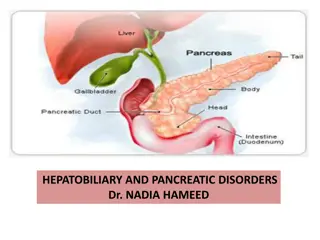
Cirrhosis of the Liver Dr .Nadia Zawi Gastroenterology BMC
objectives 1-Physiological background of the liver . 2-Description and definition of cirrhosis. 3-Specific causes, clinical picture of cirrhosis . 4- Evaluation of cirrhosis. 5 - the most common complications, and their management .
FUNCTIONS OF THE LIVER Regulating blood glucose level by making glycogen, which is stored in hepatocytes. Synthesizing blood glucose from amino acids of lactate through gluconeogenesis. Converting ammonia produced from gluconeogenetic by- products and bacteria to urea. Synthesizing plasma proteins such as albumin, globulins, clotting factors, and lipoproteins. Breaking down fatty acids into ketone bodies. Storing vitamins and trace metals. Affecting drug metabolism and detoxification. Secreting bile.
Definitionof cirrhosis Is a chronic disease characterized by scaring and necrotic tissue replaced by fibrotic tissue . Resulting in hepatic insufficiency and portal hypertension
causes 1- Chronic alcoholic abuse. 2- Non alcoholic steatohepatitis. 3- Chronic viral infection(HBV,HCV). 4- Autoimmune : BPS .PSC,AIH. 5- Genetic disorders: heamochromatosis, alfa 1 antitrypsine deficiency,wilsons disease. 6- Buddchiari syndrome. 7- Drugs: amiodarome , methyldopa, methotrexate.
Clinical Manifestations Onset usually insidious GI disturbances: Anorexia Dyspepsia Flatulence N-V, change in bowel habits Abdominal pain. Fever Lassitude (laziness) Weight loss Fetor hepaticus Jaundice Asterixis Parotid gland enlargement Hepatomegaly Splenomegaly Caput medusa
Laboratory Studies most common measured laboratory test classified as LFTs include the enzyme tests (principally the serum aminotransferases (AST &ALT), alkaline phosphatase, and gamma glutamyl transpeptidase), the serum bilirubin tests of synthetic function (principally the serum albumin concentration and prothrombin time)
Radiologic Modalities to detect complications of cirrhosis. Can occasionally suggest the presence of cirrhosis:- Uss. Ct scan. Fibroscan is more sensitive in detecting the degree of fibrosis , eves if uss was normal. Esophagoscopy to assess varices
Normal liver Cirrhotic liver
Liver biopsy Sensitivity of a liver biopsy for cirrhosis is in the range of 80 to 100. not necessary if the clinical, laboratory, and radiologic data strongly suggest the presence of cirrhosis. liver biopsy can reveal the underlying cause of cirrhosis.
Evaluation of Cirrhosis No treatment can reverse cirrhosis, or ensure that no further progression occurs. Medical therapy improve general health and alleviate symptoms . The main objectives are to 1-Detect treatable causes. 2-Prevent and correct malnutrition. 3- Treat complications. a specific etiology can usually be determined by the history combined with serologic and histologic evaluation
Evaluation of Cirrhosis Stop alcohol and toxic drugs. Correct nutritional status by vitamins, nutritional supplements high calorie and low protein. Treat ascities and fluid over load. Treatment of other problems associated with liver failure such as anti emetic , lactulose, and streptomycin for hepatic encephalopathy. esophageal varices. Liver transplantation .
Regular follow up Clinical +LFT+CBC every 3 months. US + AFP 6 months. Upper Endoscopy 1-2 years. General check up every year. (Bl. sugar, KFT, Urine, etc)
Complications of liver cirrhosis Ascites Spontaneous Bacterial Peritonitis Hepatorenal syndrome Variceal hemorrhage Hepatopulmonary syndrome Other Pulmonary syndromes Hepatic hydrothorax Portopulmonary HTN Hepatic Encephalopathy Hepatocellular carcinoma
Ascites Accumulation of fluid within the peritoneal cavity, as a result of portal HTN . Most common complication of cirrhosis . Other causes of ascites:- o heart failure o renal failure o pancreatic disease o tuberculosis. o cancer . o Hypothyroidism .
Management of ascites Aim is to relieve dicomfort , or detect other causes. Assess liver function. Evaluation of renal and CVS function. Ascitic fluid analysis. Imaging study which include: Ultrasound with Dopplers Easily confirms ascites May see nodularity of cirrhosis Evaluate patency of vasculature CT / MRI Evaluation for malignancy
Cell count: Greater than 250 PMN = Spontaneous bacterial peritonitis( SBP). If ascites is bloody ( > 50,000 RBC/mm3). bloody Non-traumatic ascitis associated with malignancy. Serum to Ascites Albumin Gradient: it is superior to the exudate-transudate concept in the differential diagnosis of ascites. (Serum albumin ascitic albumin) 97% accurate SAAG > 1.1 g/dL Portal HTN SAAG < 1.1 g/dL Other causes
Total Protein. ( > 2.5 g/dL) : TB, malignancy , hepatic venous obstruction , pancreatic ascitis . Glucose. Infection and cancer consume glucose low LDH. Ascitis/Serum LDH ratio ~ 0.4 in cirrhotic ascites Approaches 1.0 in SBP >1.0, usually infection or tumor
Amylase Uncomplicated cirrhotic ascites About 40 IU/L. The AF/S ratio is about 0.4 Pancreatic ascites About 2000 IU/L. The AF/S ratio is about 6 Triglycerides run on milky fluid. Chylous ascites - TG > 200 mg/dL, usually 1000 mg/dL Bilirubin run on brown ascites. Biliary perforation AF Bili > serum Bili
Management of ascites Initial therapy: Na restriction( low salt diet). Diuretics: spironolactone (100-400mg) Frusemide(40mg) (stepwise increase as needed to maximal doses). weight and assess abd girth . Therapeutic paracentesis Removing up to 5L appears safe. No good data on role of volume expanders( iv human aibumin). Peritoneovenous shunts.
Spontaneous Bacterial Peritonitis Infection of ascitic fluid Almost always seen in the setting of end-stage liver disease The diagnosis is established by:- A positive ascitic fluid bacterial culture ( Ecoli,klebsiell, strept) Elevated ascitic fluid absolute polymorphonuclear leukocyte (PMN) count ( >250 cells/mm3). Clinical manifestations: Fever , Abdominal pain , Altered mental status.
Cefotaxime or tazocin are effective for 5 days or until sensitivies known. Prophylactic norfloxacin 400mg is recommended for high risk patients low albumin. prolonged INR. low ascitic albumin. Previous episode of SBP.
Oesophageal varices Tortuous, dilated and engorged oesophageal veins, seen along the longitudinal axis of oesophagus. Elevated pressure in the portal venous system in liver cirrhosis. Bypassing of portal venous blood from the liver to oesophageal venous plexus. Manifested clinically by : haematemsis, melena , or fresh rectal bleeding .
Variceal hemorrhage Occurs in 25 to 40 percent of patients with cirrhosis Prophylactic measures( b.Blockers, nitrates) that redce portal vein pressure. Screening EGD recommended for all cirrhotic patients. Endoscopic band ligation is indicated for advanced or bleeding varices. Injection of vasopressins.
Oesophageal varices Band ligation
prevention and treatment principles of variceal hemorrhage: Prediction of patients at risk . Prophylaxis against a first bleed . Treatment of an active bleed . Prevention of Rebleeding . Risk factors for variceal bleeding : 1- pressure within the varix. 2- variceal size. 3- tension on the variceal wall. 4-severity of liver disease.
Hepatorenal syndrome acute renal failure coupled with advanced hepatic disease (due to cirrhosis or less often metastatic tumor or severe alcoholic hepatitis) in absence of an identifiable cause of renal failure. characterized by: Oliguria benign urine sediment very low rate of sodium excretion progressive rise in the plasma creatinine concentration( no improvement with treatment).
Hepatorenal syndrome Reduction in GFR often clinically masked No current use of Nephrotoxic agents confirms the diagnosis of HRS. Prognosis is poor unless hepatic function improves.
Hepatic Hydrothorax Pleural effusion in a patient with cirrhosis and no evidence of underlying cardiopulmonary disease is usually right-sided Diagnosis -pleural fluid analysis reveals a transudative fluid serum to fluid albumin gradient greater than 1.1 Treatment with : Diuretics(frusemide). Pleural fluid aspiration indicated if patient developed respiratory emparssement.
Hepatic Encephalopathy Spectrum of potentially reversible neuropsychiatric abnormalities seen in patients with liver dysfunction Diurnal sleep pattern pertubation Asterixis Hyperactive deep tendon reflexes Transient decerebrate posturing
Hepatic Encephalopathy Monitoring for events likely to precipitate HE [i.e.- variceal bleeding, infection (such as SBP), the administration of sedatives, hypokalemia, and hyponatremia] Reduction of ammoniagenic substrates Lactulose / lactitol Dietary restriction of protein Zinc and melatonin
Hepatocellular Carcinoma Patients with cirrhosis have a markedly increased risk of developing hepatocellular carcinoma Incidence in well compensated cirrhosis is approximately 3 percent per year Symptoms are largely due to mass effect from the tumor Pain, early satiety, obstructive jaundice, and a palpable mass Serum AFP greater than 500 micrograms/l in a patient with cirrhosis are virtually diagnostic Median survival following diagnosis is approximately 6 to 20 months
Treatment Options of liver cirrhosis SUMMARY The major goals of treating the cirrhotic patient include: Slowing or reversing the progression of liver disease directed to under lying cause. Preventing superimposed insults to the liver. Preventing and treating the complications. Determining the appropriateness and optimal timing for liver transplantation .
Liver Transplantation Liver transplantation is the definitive treatment for patients with decompensated cirrhosis Depends upon the severity of disease, quality of life and the absence of contraindications