Understanding Erythema Multiforme: Symptoms, Causes & Treatment
Learn about erythema multiforme (EM), a dermatitis condition characterized by distinctive skin lesions. Discover the types, causes, clinical features, and more about this condition that primarily affects young adults.

Download Presentation

Please find below an Image/Link to download the presentation.
The content on the website is provided AS IS for your information and personal use only. It may not be sold, licensed, or shared on other websites without obtaining consent from the author. If you encounter any issues during the download, it is possible that the publisher has removed the file from their server.
You are allowed to download the files provided on this website for personal or commercial use, subject to the condition that they are used lawfully. All files are the property of their respective owners.
The content on the website is provided AS IS for your information and personal use only. It may not be sold, licensed, or shared on other websites without obtaining consent from the author.
E N D
Presentation Transcript
Erythema Multiforme Stevens-Johnsonsyndrome, erythema multiformemajor, Erythema multiforme minor, herpes-induced EMmajor, herpes-associated erythemamultiforme, drug-induced Stevens-Johnsonsyndrome
Introduction An acute self-limiting dermatitis characterized by a distinctive clinical eruption manifested as the iris ortarget lesion. Types of EM: EMminor:localized eruption of the skinwith mild or no mucosalinvolvement EMmajor skin and mucosal erosions of raised atypical targetlesions, usuallylocatedonthe extremities and/or onthe face. Stevens-Johnson syndrome(SJS) skin and mucosal erosions plus widespread distribution of flatatypical targets or purpuricmacules, maybepresentonthe trunk, the face,andonthe extremities. SJS is considered under separate disease now etiopathogenetically
Etiology EM and SJS are both caused by drugs, butinfectious agents are considered tobe the major cause of EM. History of HSV infection 1 to 3 weeks beforeonset of EM EM minor: triggered by HSV in nearly 100% ofcases EM major: herpetic etiology also accounts for55% of cases of, other infections,Mycoplasma S J S and EM major: Drugs are found to be major cause, antibacterial sulfonamides,anticonvulsants, oxicam, NSAIDs, and allopurinol
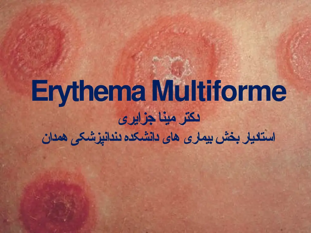
ClinicalFeatures Chiefly in young adults, although itmay develop at anyage, Second to fourth decades oflife Affects males more frequently thanfemales. Characterized by the occurrenceof asymptomatic, vividly erythematous discrete macules,papules occasionally vesicles and bullae distributed in arather symmetrical pattern most commonly over the hands and arms, feet and legs, face andneck. Variable size, but are generally only afew centimeters or less indiameter.
ClinicalFeatures A concentric ring like appearance of the lesions, resulting from the varying shades of erythema, occurs in somecases and has given rise to the terms target , iris , or bull seye Most common on the hands, wrists andankles. Mucous membrane involvement, including the oralcavity, iscommon. The lesions make their appearancerapidly, usually within a day or two, and persist from several daysto a few weeks, gradually fading and eventuallyclearing. Recurrence of the disease over a period of years iscommon
Oral Manifestation ofEM Usually not a significant feature of thedisease Except for the pain and discomfort theycause. Hyperemic macules, papules or vesicles may become eroded or ulcerated and bleedfreely. The tongue, palate, buccal mucosa and gingiva are commonly diffuselyinvolved Occasionally, mucous membrane lesionsoccur before the cutaneous manifestations, but oral involvement without dermal lesions has been questioned
Stevens-Johnsonsyndrome At one time considered tobe a separate disease Mucocutaneous-oculardisease Now recognized as simply a very severe bullous form of EM With widespread involvement typically includingthe skin, oral cavity, eyes andgenitalia. It commences with the abruptoccurrenceof fever, malaise, photophobia, and eruptions of the oral mucosa, genitalia andskin. Thecutaneouslesionsin this aresimilarto thoseof EM, Although they are commonly hemorrhagicand are often vesicular orbullous
ClinicalFeatures Eyelesions photophobia, a characteristic of the disease referable tothe conjunctivitis, corneal ulceration and panophthalmitis which mayoccur Keratoconjunctivitis sicca also has beendescribed. Blindness may result chiefly from intercurrent bacterialinfection. Genitallesions Nonspecificurethritis, balanitis and/or vaginalulcers Other reported complications are related to respiratory tract involvement such as tracheobronchial ulcerationand pneumonia. The patients usually recover unless they succumb toa secondaryinfection.
Oral Manifestation ofS J S Extremely severe and so painful thatmastication isimpossible. Mucosal vesicles or bullae occur which rupture and leave surfaces covered with a thick whiteor yellowexudate. Erosions of the pharynx are alsocommon The lips may exhibit ulceration withbloody crusting and arepainful
Oral Manifestation ofS J S May bethe chief complaint of the patient, and understandably, have been mistaken for acute necrotizingulcerativegingivostomatitis. Interestingly, however, it has been reported that the organisms of Vincent s infectionare scarce in patients with thisdisease. The mucosal involvement in SJS is more severe and extensive than in EMmajor
HistologicFeatures Not of diagnosticimportance. Although considerable variation occurs, corresponding to the variation in clinical appearance Cutaneous or mucosal lesions generallyexhibit intracellular edema of the spinous layer of epithelium Edema of the superficial connective tissuewhich may actually produce a subepidermalvesicle. Zone of severeliquefactiondegeneration - upper layers of theepithelium,
HistologicFeatures Intraepithelial vesicle formation andthinning with frequent absence of the basement membrane. Dilatation of the superficial capillariesand lymphaticvessels Varying degree of inflammatory cell infiltration, chiefly lymphocytes, but often neutrophils and eosinophils, is alsopresent
DifferentialDiagnosis Varied nature of the disease - difficulty in diagnosis, particularly when the occurrence ofcutaneous lesions isminimal. In the presence of orallesions, Aphthousstomatitis, Contact dermatitis orstomatitis Acute necrotizinggingivitis Pemphigus, Dermatitisherpetiformis, Bullous lichenplanus, herpeszoster, chickenpox Toxic epidermal necrolysis (Lyell sdisease)
Toxic Epidermal Necrolysis(TENS) very serious, often fatal, bullous drugeruption So severe that large sheets of skin peel off- appearance of a widespread scaldingburn. Oral erosions may alsooccur Considered to be a confluent form ofStevens- Johnsonsyndrome. TENS must be differentiated fromthe Staphylococcal Scalded Skin Syndrome (SSS) clinically similar even though the latter is amilder disease with a betterprognosis
TREATMENT Identificationof the causeshouldbemade if possible. If a drug is suspected, itmust be withdrawn. Infections should be appropriately treatedafter cultures and/or serologictests For all forms of EM symptomatictreatment Oral antihistamines,analgesics, Local skin care: liquid antiseptics 0.05%chlorhexidine soothing mouthwashes Topical steroids may beconsidered. Oral antacids - discrete oralulcers. S J S and TENS: treatment is generally with high doses of systemic corticosteroids, intravenous immunoglobulin,and thalidomide
Refrences Shafers, Oral Pathology 6thedition Burket s ORAL MEDICINE 11thEdition Regezi: Oral Pathology: Clinical Pathologic Correlations, 5th ed & 6thedition Essential of Oral Pathology and medicine 7th ed: Cawsons & odell Color atlas of Oral Pathology:Nevile Pathology of the Head and Neck:Antonio Cardesa, Pieter J.Slootweg Essential of Oral Pathology : SwapanKumar Purkait
THANKS www.facebook.com/notesdental www.facebook.com/notesdental


")
")


