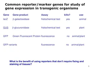
Understanding Fluorescence Technology in Biology
Explore the applications and advantages of fluorescence technology in biology, including the use of fluorescent protein reporters, fluorescence microscopy, and tagging target molecules for in vivo detection. Learn how fluorescence is used in various biological processes such as cellular integrity, DNA sequencing, and genetic mapping. Discover the benefits of fluorescence technology for imaging techniques and identifying multiple target molecules simultaneously.

Download Presentation

Please find below an Image/Link to download the presentation.
The content on the website is provided AS IS for your information and personal use only. It may not be sold, licensed, or shared on other websites without obtaining consent from the author. If you encounter any issues during the download, it is possible that the publisher has removed the file from their server.
You are allowed to download the files provided on this website for personal or commercial use, subject to the condition that they are used lawfully. All files are the property of their respective owners.
The content on the website is provided AS IS for your information and personal use only. It may not be sold, licensed, or shared on other websites without obtaining consent from the author.
E N D
Presentation Transcript
Fluorescent Protein Reporters and Fluorescence Technology Josh Leung James Weis February 18th, 2010 Bio 1220, Gary Wessel
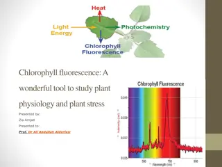
Fluorescence: How does it work? Fluorescence vs Phosphorescence -Time delay microsecond vs min Photon absorbed photon released -One photon vs two photons GFP, other technologies mainly use fluorescence Jablonski Diagram
What is Fluorescence used for? In Biology Fluorescent proteins and fluorophore tagging cellular integrity, endocytosis, exocytosis, membrane fluidity, protein trafficking, signal transduction, enzymatic activity, genetic mapping, etc. Fluorescence Microscopy DNA Microarrays (test for gene expression) DNA Sequencing Fluorescence Recovery After Photobleaching Fluorescence-Activated Cell Sorting Other uses include fluorescent lighting, flame tests, etc.
Advantages of Fluorescence Technology Tagging a target molecule In vivo detection Reliable (even down to one molecule) High fidelity and specificity Identify multiple target molecules simultaneously Development of new imaging techniques Also detect more types of targets
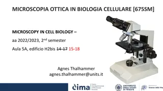
Fluorescence Microscopy Shine light fluorescence detection Separate weaker fluorescence from the excitation light using filters Limit of detection determined by the darkness of the background (lack of noise, etc) Camera Specimen
C. Elegans Nervous System Cell Division Fluorescence Microscope
Endothelium Cells (Triple fluorescence staining of endothelium cells from a pulmonary artery) Mammalian Cells (DNA is blue, microfilaments are green) Fluorescence Microscope
Opossum Kidney Cortex Epithelial Cells (OK Line)
Human Cervical Adenocarcinoma Cells (HeLa Line)
FRAP Fluorescence recovery after photo-bleaching Study diffusion and movement of biological molecules fluid mosaic model of the cell membrane study molecules in the cytosol, nucleus, etc Fluorescence Time
Fluorescence-Activated Cell Sorting Rapid sorting Sorts cells one-by-one
Microarrays DNA (Gene) microarrays Gene expression profiling (using fluorescent labeled mRNA) SNP detection Protein microarrays Antibody analysis Protein interactions
Reporter Genes Attached to genes of interest Chosen by the characteristics they confer to the organism expressing them Easily identified / measured Selectable markers Determine whether the gene of interest is being expressed
Common uses of reporter genes Gene expression assays Promoter assays Transformation / transfection assays Two-hybrid screening
So, what makes a good reporter gene? Genes that confer easily identifiable characteristics. Green Fluorescent Protein (GFP) Jellyfish Causes cells to glow green under blue light Red Fluorescent Protein (dsRed) Coral Luciferase Fireflys Catalyzes a reaction with luciferin, producing light
GFP Aequorea victoria 238 amino acids Refined from WT over the years 1995; Mutation dramatically improving the spectral characteristics of GFP 1995; F64L, allowing GFP use in mammalian cells Variants Superfolder GFP Blue, Cyan, Yellow, Red, Emerald, Apple
Fluorescent proteins and their uses Fluorescent proteins derived from GFP and dsRed. Colors: BFP mTFP1 Emerald Citrine mOrange mApple mCherry mGrape
Florescent proteins Fluorescence microscopy Florescent proteins not phototoxic, as are most florescent molecules Determine when gene is expressed Exhibit morphological distinctions View biological processes (protein folding, transport, etc) Expression of a florescent protein in specific cells Optical detection of specific cells
Two color male pig kidney epithelial cells undergoing mitosis A culture of pig kidney cells mCherry fused to human histone H2B mEmerald fused to alpha-tubulin