Understanding Imaging for Head Injuries
Learn about the importance of imaging in diagnosing head injuries, including traumatic brain injuries, concussions, and subdural hematomas. Discover real-life cases where early imaging made a critical difference in patient outcomes.
Download Presentation

Please find below an Image/Link to download the presentation.
The content on the website is provided AS IS for your information and personal use only. It may not be sold, licensed, or shared on other websites without obtaining consent from the author. If you encounter any issues during the download, it is possible that the publisher has removed the file from their server.
You are allowed to download the files provided on this website for personal or commercial use, subject to the condition that they are used lawfully. All files are the property of their respective owners.
The content on the website is provided AS IS for your information and personal use only. It may not be sold, licensed, or shared on other websites without obtaining consent from the author.
E N D
Presentation Transcript
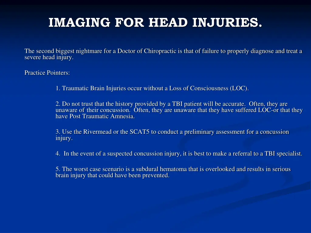
IMAGING FOR HEAD INJURIES. The second biggest nightmare for a Doctor of Chiropractic is that of failure to properly diagnose and treat a severe head injury. Practice Pointers: 1. Traumatic Brain Injuries occur without a Loss of Consciousness (LOC). 2. Do not trust that the history provided by a TBI patient will be accurate. Often, they are unaware of their concussion. Often, they are unaware that they have suffered LOC-or that they have Post Traumatic Amnesia. 3. Use the Rivermead or the SCAT5 to conduct a preliminary assessment for a concussion injury. 4. In the event of a suspected concussion injury, it is best to make a referral to a TBI specialist. 5. The worst case scenario is a subdural hematoma that is overlooked and results in serious brain injury that could have been prevented.
Status of Imaging For Traumatic Brain Injury. Currently, there is no imaging that can reliable rule out a Traumatic Brain Injury of any degree of severity (including concussions). Imaging, both CT scans and MRIs can rule in a brain hematoma. It should be remembered that brain hematomas can develop after as much as 7-10 days later after a head trauma with minimal initial symptoms that would be a red flag for a serious brain injury.
IMAGING FOR HEAD INJURIES. MRIs or CT scans will rule out a subdural hematoma in almost all cases-and protect the patient and protect you from a medical malpractice claim. I have had two cases in which the subdural hematoma was overlooked and developed later.
IMAGING FOR HEAD INJURIES. Jacob s Case: Jacob was partying with friends and probably had too much to drink. He slipped on ice and banged his head hard on concrete. The ER doctor failed to perform any imaging. He experienced headaches, turning severe almost 10 days later. Imaging detected a subdural hematoma, resulting in largely unsuccessful surgery. The result was serious permanent brain damage. He has been declared totally disabled for life.
IMAGING FOR HEAD INJURIES. Bob s Case: Bob was on a hunting trip in the Dakota s when he slipped and fell, banging his head hard on concrete steps. Other than a scalp contusion and short term headache and dizziness, he suffered only minimal concussion symptoms for a short time. One week later, Bob experienced a rather sudden onset of severe, pounding headaches for which he sought care. Imaging revealed a subdural hematoma which resulted in successful surgery with no apparent long-term disability.
RADIOLOGY-Definitional Confusion. Standardization of language is difficult, especially among those who have expert knowledge of the subject and clear understanding of what their own words mean. The difficulties must be overcome, because deleterious effects ensue when we do not understand what one another's words mean. Existing dictionary definitions and previous efforts by experts have lacked the attention to detail and multidisciplinary consensus we brought to this work. The North American Spine Society (NASS) initiated efforts to develop detailed definitions of lumbar disc pathology terms and has provided sustained support of the project. Independent efforts by neuroradiologists led the American Society of Spine Radiology (ASSR) and American Society of Neuroradiology (ASNR) to organize a task force of neuroradiologists and encourage liaison with the NASS group. The results are this document and improved communications between the societies.
RADIOLOGY Task Force: American Academy of Orthopaedic Surgeons (AAOS) American Academy of Physical Medicine and Rehabilitation (AAPM&R) American College of Radiology (ACR) American Society of Neuroradiology (ASNR) American Society of Spine Radiology (ASSR) Joint Section on Disorders of the Spine and Peripheral Nerves of the American Association of Neurological Surgeons (AANS) and Congress of Neurological Surgeons (CNS) European Society of Neuroradiology (ESNR)
RADIOLOGY This s the statement of the Joint Study regarding the need for clarity in disc injury conditions: Physicians need reliable terms that describe normal and pathologic conditions of lumbar discs. Terms that can be interpreted accurately, consistently, and with reasonable precision are particularly important for communicating impressions gained from imaging for clinical diagnostic and therapeutic decision making. Although clear understanding of disc terminology between radiologists and clinicians is the focus of this work, such understanding can be critical, also, to patients, families, employers, insurers, jurists, social planners, and researchers.
RADIOLOGY Anular tears, also properly called anular fissures, are separations between anular fibers, avulsion of fibers from their vertebral body insertions, or breaks through fibers that extend radially, transversely, or concentrically, involving one or many layers of the anular lamellae. The terms "tear" or "fissure" describe the spectrum of such lesions and do not imply that the lesion is consequent to trauma. (Figure 2)
RADIOLOGY Herniated discs may take the form of protrusion or extrusion, based on the shape of the displaced material. Protrusion is present, if the greatest distance, in any plane, between the edges of the disc material beyond the disc space is less than the distance between the edges of the base in the same plane. The base is defined as the cross- sectional area of disc material at the outer margin of the disc space of origin, where disc material displaced beyond the disc space is continuous with disc material within the disc space. In the cranio-caudal direction, the length of the base cannot exceed, by definition, the height of the intervertebral space. Extrusion is present when, in at least one plane, any one distance between the edges of the disc material beyond the disc space is greater than the distance between the edges of the base in the same plane, or when no continuity exists between the disc material beyond the disc space and that within the disc space (Figure 12). Extrusion may be further specified as sequestration, if the displaced disc material has lost completely any continuity with the parent disc. Migration signifies displacement of disc material away from the site of extrusion, regardless of whether sequestrated or not. Because posteriorly displaced disc material is often constrained by the posterior longitudinal ligament, images may portray a disc displacement as a protrusion on axial sections and an extrusion on sagittal sections, in which cases the displacement should be considered an extrusion. Herniated discs in the cranio-caudal (vertical) direction through a break in the vertebral body end-plate are referred to as intravertebral herniations .
Herniation Herniation is defined as a localized displacement of disc material beyond the limits of the intervertebral disc space term "localized" contrasts to "generalized , the latter being arbitrarily defined as greater than 50% (180 degrees) of the periphery of the disc being displaced from the disc space (defined craniad and caudad by the vertebral ring apophyses).
Focal means localized displacement in the axial (horizontal) plane of less than 25% of the disc circumference. Broad Based means that between 25 and 50% of the disc circumference is displaced. Bulging is the presence of disc tissue Circumferentially" (50-100%) beyond the edges of the ring apophyses. (not considered a form of herniation).
DEFINITIONS OF HERNIATION. Herniated discs may take the form of protrusion or extrusion, based on the shape of the displaced material. Protrusion is present, if the greatest distance, in any plane, between the edges of the disc material beyond the disc space is less than the distance between the edges of the base in the same plane. The base is defined as the cross-sectional area of disc material at the outer margin of the disc space of origin, where disc material displaced beyond the disc space is continuous with disc material within the disc space. In the cranio-caudal direction, the length of the base cannot exceed, by definition, the height of the interertebral space.
Extrusion is present when, in at least one plane, any one distance between the edges of the disc material beyond the disc space is greater than the distance between the edges of the base in the same plane, or when no continuity exists between the disc material beyond the disc space and that within the disc space. Sequestration exists if the displaced disc material has lost completely any continuity with the parent disc. Migration signifies displacement of disc material away from the site of Extrusion, regardless of whether sequestrated or not.
Because posteriorly displaced disc material is often constrained by the posterior longitudinal ligament, images may portray a disc displacement as a protrusion on axial sections and an extrusion on sagittal sections, in which cases the displacement should be considered an extrusion on sagittal sections, in which cases the displacement should be considered an extrusion. Contained means that the displaced portion is covered by outer anulus, or uncontained when absent any such covering. Displaced disc tissues may also be described by location, volume, and content. Figure 15 lists the proposed categories for description and classification of disc herniations.

