Explore the composition of skeletal tissue and muscle fibers, learn about contractile proteins essential for muscle function, and delve into the organization of the contractile unit in muscles. Discover the intricate details of muscle anatomy and physiology.
Please find below an Image/Link to download the presentation.
The content on the website is provided AS IS for your information and personal use only. It may not be sold, licensed, or shared on other websites without obtaining consent from the author. If you encounter any issues during the download, it is possible that the publisher has removed the file from their server.
You are allowed to download the files provided on this website for personal or commercial use, subject to the condition that they are used lawfully. All files are the property of their respective owners.
The content on the website is provided AS IS for your information and personal use only. It may not be sold, licensed, or shared on other websites without obtaining consent from the author.
E N D
Presentation Transcript
Dr Payal Dhawale Dept. Of Sports Physiotherapy MGM Institute Of Physiotherapy Chh. Sambhajinagar
Skeletal tissue (noncontractile). muscles (contractile) are composed and of Muscle tissue connective Muscle tissue has the ability to develop tension in mechanical stimuli. response to chemical, electrical, or The connective tissue, on the other hand, develops loading. tension in response to passive
Contractile Proteins A skeletal muscle is composed of many thousands of muscle fibers. Contractile Proteins A single muscle contains many fascicles, muscle fibers (cells) surrounded by connective tissue. fascicles, a group of Each fiber is a single muscle cell that is enclosed in a cell membrane called the sarcolemma sarcolemma Like other cells in the body, the muscle fiber is composed of cytoplasm, which in a muscle is called sarcoplasm sarcoplasm. .
The contractile structures of a muscle fiber and nonmyofibrillar structures such as ribosomes, glycogen, and mitochondria, which are required for cell metabolism. The sarcoplasm sarcoplasm contains myofibrils contains myofibrils which are the The myofibril is composed of thick myofilaments composed of the protein myosin and thin filaments composed of the protein actin myofilaments myosin and thin filaments actin. . The essential for a muscle contraction to occur. The interaction of these two myofilaments is
Organization of the Contractile Unit The portion of the myofibril that is located between two Z disks is called the sarcomere Organization of the Contractile Unit sarcomere. . The Z throughout the myofibril, not only serve as boundaries for the sarcomere but also link the thin filaments together. The Z disks, which are located at regular intervals Areas of the sarcomere called bands identify bands or zones help to zones help to identify the arrangement of the thick and thin filaments. The portion of the sarcomere that extends over both the length of the thick filaments and a small portion of the thin filaments is called the anisotropic or A band. anisotropic or A band.
Areas that include only actin filaments are called isotropic or I bands. isotropic or I bands. The terms anisotropic and isotropic refer to the behavior of these portions of the fibers when light shines on them. The central portion of the thick filament (A band area) in which there is no overlap with the thin filaments is called the H zone. H zone. The central portion of the H zone, which consists of the wide middle portion of the thick filament, is called the M band. M band.
Interaction between the thick and thin filaments of the sarcomere leading to muscle contraction is initiated by the arrival of a nerve impulse at the motor end plate, which evokes an electric impulse, or action potential, that travels along the muscle fiber.
The action potential initiates the release of calcium ions, which cause troponin to reposition the tropomyosin molecules so that receptor sites on the actin are free and the head groups of the myosin can bind with actin. This bonding of filaments is called a cross-bridge
Organization Organization of of the the Motor Motor Unit Unit Although the sarcomere is the basic unit of tension in a muscle, it is actually part of a larger complex called the motor unit. The motor neuron and all of the muscle fibers it innervates. motor unit unit consists consists of of the the alpha alpha motor motor The stimulus that the muscle fiber receives initiating the contractile process is transmitted through an alpha alpha motor motor neuron neuron. .
The contraction of the entire muscle is the result of many motor units firing asynchronously and repeatedly. The magnitude of the contraction of the entire muscle may be altered by changing the number of motor units that are activated or the frequency at which they are activated. The number of motor units in a muscle, as well as the structure of these units, varies from muscle to muscle.
This recruitment strategy is referred to as the size principle of motor unit recruitment. Small motor units generate less tension than do large motor units and require less energy expenditure, and therefore this recruitment strategy is thought to be energy conserving. If a few small motor units are capable of accomplishing the task, the recruitment of large motor units is unnecessary. If the task demands are such that the small motor units are unable to complete the task, larger motor units can be recruited.
However, the recruitment strategy may be based not only on energy conservation but also on previous experience: Nature of the task (how rapidly the muscle must respond or the anticipated magnitude of the required force) Type of muscle action (concentric, eccentric, or isometric) Mechanism that takes into account the actions of all muscles around a joint, including such considerations as the muscle s mechanical advantage at a particular point in the range of motion (ROM).
Fiber Types Fiber Types - Type I (slow) - Type IIA (intermediate) - Type IIB(fast)
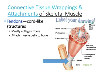
Organization of Connective Tissue in Muscle The sarcolemma of individual muscle fibers is surrounded by connective tissue called the endomysium groups of connective tissue called the perimysium The myofibril is connected to the via specialized proteins. The endomysium and perimysium are continuous with the outer connective tissue sheath called the epimysium endomysium, , groups of muscle fibers are covered by perimysium. . endomysium The myofibril is connected to the endomysium epimysium, which envelops the entire muscle. , which envelops the entire muscle.
The zone of superficial loose dermis. This zone contributes to the mobility of the skin, acts as an insulator, and may contain skin muscles such as the platysma in the neck. superficial fasciae, tissue, is is located fasciae, composed located directly under the composed of of loose tissue,
The zone of deep compacted fibers. The deep fasciae attach to muscles and bones and may form tracts or bands and retinacula. For example, the deep femoral fasciae in the lower extremity forms a tract known as the iliotibial This lowerextremity and the gluteus maximus) to the bones of the leg. deep fasciae and regularly fasciae is is composed regularly arranged collagenous composed of of compacted and iliotibial tract This tract lowerextremity muscles (the tensor fascia latae tract or tract transmits or band transmits the band. . the pull pull of of two two of of the the
Retinacula thickenings of the fasciae, which form a loop that is attached at both ends to bone. The tunnels formed by retinacula retain or prevent tendons from bowing out of position during muscle action . Retinacula are formed by localized transverse
All of the connective tissue in a muscle is interconnected and constitutes the passive elastic The connective tissues that surround the muscle, plus the sarcolemma, the elastic protein titin (connectin), and other structures (i.e., nerves and blood vessels), form the parallel passive elastic component component of of a a muscle muscle. . parallel elastic elastic component component of of a a muscle muscle. .
When sarcomeres shorten from their resting positions, the slack collagen fibers within the parallel elastic component buckle even further. Whatever tension might have existed in the collagen at rest is diminished by the shortening of the sarcomere. Because components of a muscle, the increase or decrease in passive tension can substantially affect the total tension output of a muscle. buckle (crimp) (crimp) of the many parallel elastic
The tendon of the muscle is considered to function in series elements This tension when the muscle actively produces tension. When the contractile elements in a muscle actively shorten, they exert a pull on the tendon series with with the the contractile contractile elements. . This means means that that the the tendon will be under
Thus, most of the muscle force can be used for moving the bony lever and is not dissipated stretching the tendon. The tendon is also under tension when a muscle is controlling or braking the motion of the lever in an eccentric contraction. A tendon is under reduced tension only when a muscle is completely relaxed and in a relatively shortened position.
Muscle Tension The most important characteristic of a muscle is its ability to develop tension and to exert a force on the bony lever. Tension can be either active or passive, and the total tension that a muscle can develop includes both Active and passive components.
Passive tension refers to tension developed in the parallel elastic component of the muscle. The parallel elastic component may add to the active tension produced by the muscle when the muscle is lengthened, or it may become slack and not contribute to the total tension when the muscle is shortened. The total tension that develops during an active contraction of a muscle is a combination of the passive (noncontractile) tension added to the active (contractile) tension.
Active tension refers to tension developed by the contractile elements of the muscle. Active tension in a muscle is initiated by cross-bridge formation and movement of the thick and thin filaments. The amount of active tension that a muscle can generate depends on neural factors and mechanical properties of the muscle fibers.
The neural factors that can modulate the amount of active tension include the frequency, number, and size of motor units that are firing. The mechanical properties of muscle that determine the active tension are the isometric length-tension relationship and the force velocity relationship.
One of the most fundamental concepts in muscle physiology is the direct relationship between isometric tension development in a muscle fiber and the length of the sarcomere in a muscle fiber.
Muscle fibers develop maximal isometric tension at optimal sarcomere length because the thick and thin filaments are positioned so that the maximum number of cross-bridges within the sarcomere can be formed.
If the muscle fiber is lengthened or shortened beyond optimal length, the amount of active tension that the muscle fiber is able to generate when stimulated decreases. When a muscle fiber is lengthened beyond optimal length, there is less overlap between the thick and thin filaments and consequently fewer formation. possibilities for cross-bridge
the passive elastic tension in the parallel component may be increased when the muscle is elongated. This passive tension is added to the active tension, resulting in the total tension
It must be remembered that the sarcomere lengthtension relationship was determined with isometric contractions and therefore should apply, in the strict sense, only to isometric muscle contraction.
During dynamic contractions, the length- tension relationship must be combined with the force-velocity relationship to determine the effect that both length and velocity have on muscle tension.