Understanding Regional Circulation and Coronary Blood Flow
Explore the concept of regional circulation which involves the regulation of blood flow in specific body regions based on local activity levels. Learn about the physiological importance of various regional circulations such as coronary, cerebral, and pulmonary circulation. Dive into the details of coronary circulation, including the arteries, venous drainage, and factors regulating blood flow. Gain insights into the significance of physiological shunts and the normal coronary blood flow rate.

Download Presentation

Please find below an Image/Link to download the presentation.
The content on the website is provided AS IS for your information and personal use only. It may not be sold, licensed, or shared on other websites without obtaining consent from the author. If you encounter any issues during the download, it is possible that the publisher has removed the file from their server.
You are allowed to download the files provided on this website for personal or commercial use, subject to the condition that they are used lawfully. All files are the property of their respective owners.
The content on the website is provided AS IS for your information and personal use only. It may not be sold, licensed, or shared on other websites without obtaining consent from the author.
E N D
Presentation Transcript
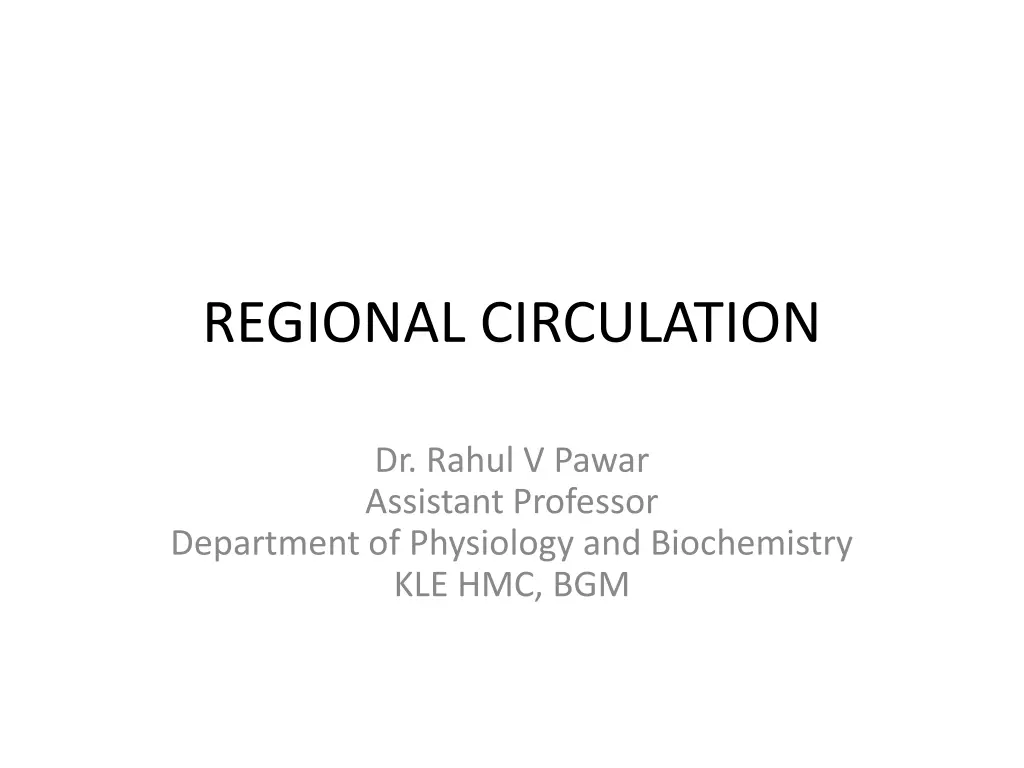
REGIONAL CIRCULATION Dr. Rahul V Pawar Assistant Professor Department of Physiology and Biochemistry KLE HMC, BGM
Regional Circulation Circulation of blood through a particular organ or a region of the body is called regional circulation. Regional circulation is directly proportional to the degree of local activity. It is achieved by: a) Regulation of general circulation, and b) Regulation of local circulation, which includes vasodilatation of the Active part and vasoconstriction of other Inactive parts. Thus shifting blood from the inactive parts to the active parts without fall in general blood pressure.
Regional Circulations of Physiological Importance: a) Coronary Circulation b) Cerebral Circulation c) Pulmonary Circulation d) Hepatic Circulation e) Splenic Circulation f) Renal Circulation g) Capillary Circulation h) Cutaneous Circulation i) Skeletal Muscle Circulation
CORONARY CIRCULATION It consists of circulation of blood through the blood vessels supplying the Myocardium. Blood flowing through the chambers of the heart does not nourish the myocardium. Coronary Blood Vessels: a) Coronary Arteries: - these include Right and Left Coronary arteries arising from the root of the Aorta. - Left coronary artery divides into Anterior Descending branch (running downward to the apex) and Left Circumflex Branch (which runs in left AV groove and runs down as Posterior Descending Branch).
b) Venous Drainage: Venous drainage from heart occurs by 3 types of vessels: 1. Coronary Sinus: Larger vein draining 75% of total coronary flow. Drains from left side of the heart into Right Atrium near Tricuspid Valve. 2. Anterior Coronary Veins: Drain blood from the Right side of the heart directly into Right Atrium. 3. Thebesian Veins: Drain deoxygenated blood from myocardium, directly into the concerned chambers of the Heart.
Physiological Shunt: - It is the diversion where the deoxygenated blood mixes with the oxygenated blood. - Physiological shunt includes 2 components: i) Drainage of deoxygenated blood from thebesian veins into the cardiac chambers. ii) Drainage of deoxygenated blood from bronchial circulation into Pulmonary Vein. Normal Coronary Blood Flow: - About 200 ml/min. - 4% of Cardiac Output - About 65-70 ml/min/100 gm of cardiac muscle.
Factors regulating Coronary Blood Flow Heart regulates its blood supply through Autoregulation Mechanism. Factors regulating: 1. Need for Oxygen: Blood passing through coronary circulation is directly proportional to O2 consumption by cardiac muscle. Hypoxia immediately causes coronary vasodilatation and increases the blood flow to the heart. 2. Metabolic Factors: Metabolites cause vasodilatation and increase coronary blood flow (Reactive Hyperemia). These include Adenosine, Potassium, Hydrogen, CO2, Adenosine phosphate compounds.
3. Coronary Perfusion Pressure: Perfusion pressure is the difference between mean arterial pressure and venous pressure. Coronary perfusion pressure is the difference between mean arterial pressure in the aorta and the right atrial pressure. This pressure is significant and is major factor for maintaining coronary blood flow. 4. Nervous Factor: Cardiac musculature and Coronary blood vessels are innervated by Sympathetic and Parasympathetic divisions of ANS. Sympathetic stimulation work done by heart more metabolites vasodilatation coronary blood flow. Parasympathetic stimulation has opposite effect.
Important Features of Coronary Circulation: Very short and very rapid Blood flow mainly occurs during diastolic phase No effective anastomoses between coronary vessels Rich circulation, 4% of cardiac output Regulated mainly by autoregulation by metabolites Capillary permeability is high, thus cardiac lymph is rich in protein Coronary vessels are highly susceptible to degeneration and atherosclerosis.
APPLIED PHYSIOLOGY CORONARY ARTERY DISEASE (CAD) The heart disease caused by inadequate blood supply to cardiac muscle due to occlusion of coronary artery. Also called Coronary Heart Disease. 1. Coronary Occlusion Partial or complete occlusion of Coronary Artery. Cause: Occlusion caused by atherosclerotic plaque (cholesterol, calcium, blood substances). This is further advanced by Thrombosis and Embolism.
Risk Factors of CAD and MI: 1. HTN 2. High level of cholesterol, triglycerides or LDL in blood 3. DM 4. Overweight and Obesity 5. Lack of physical activity 6. Smoking 7. High stress 8. Family history
2. Myocardial ischaemia and Necrosis Myocardial Ischaemia Reduced blood supply to a part of myocardium and its response to resultant hypoxia. Chief cause is occlusion of coronary artery. Ischaemia of small area of myocardium is corrected by rapid development of coronary collateral arteries. Necrosis Death of cells or tissues by injury or disease. Myocardial ischaemmia is one of the cause of necrosis. Necrosis is irreversible.
3. Myocardial Infarction Heart Attack Necrosis of myocardium caused by ischaemia due to embolus, thrombus or vascular spasm. Also called Heart Attack. Death occurs rapidly due to ventricular fibrillation. Myocardial Stunning: Transient mechanical dysfunction of the heart, caused by a mild ischaemia, not sufficient to cause necrosis. Symptoms of MI: 1. Cardiac pain Angina pectoris 2. Nausea, 3. Vomiting, 4. Palpitations, 5. Difficulty in breathing, 6. Extreme weakness, 7. Sweating, 8. Anxiety.
4. Cardiac Pain - Angina Pectoris Chest pain caused by myocardial ischaemia. Common manifestation of CAD. Pain starts beneath the sternum and radiates to the surface of left arm and left shoulder. It is a referred pain, felt away from the heart. Cause: Myocardial ischaemia accumulation of anerobic metabolic end products like uric acid, other pain producing substances like Substance P, Histamine and Kinin stimulation of sensory nerve endings sensory nerve impulses via lateral spinothalamic tract Thalamus Sensory Cortex.
CEREBRAL CIRCULATION Flow of blood through the blood vessels of Brain. Stoppage of blood flow to brain for 5 seconds unconsciousness, and for 5 minutes irreparable damage to the brain cells. Cerebral Blood Vessels: Arterial supply: Basilar artery and Internal Carotid Artery. Branches of these arteries form Circle of Willis. Venous drainage: Drained by venous sinuses which open into Internal Jugular Vein.
Normal Cerebral Blood Flow 750-800 ml/min. About 15-16 % of total Cardiac Output. About 50-55 ml/100 gm of brain tissue/min. Regulation of Cerebral Circulation Regulated by 3 factors: 1. Autoregulation: Brain regulates its blood flow by autoregulation, which depends on: i) Effective Perfusion Pressure: Venous pressure in brain is Zero, so the mean arterial pressure plays important role in regulating cerebral blood flow. Autoregulation is effective when mean arterial pressure is between 60-140 mmHg. ii) Cerebral Vascular Resistance: More the vascular resistance, less is the blood flow to the brain. Vascular resistance is offered by Intracranial Pressure, CSF pressure and Viscosity of blood.
2. Chemical Factors: which increase the cerebral blood flow are: i) Decreased O2 tension (hypoxia). ii) Increased CO2 tension. iii) Increased H-ion concentration. 3. Nervous Factors: Cerebral blood vessels are supplied by Sympathetic Vasoconstrictor Fibres. These do not play a role during normal conditions. But in HTN, they cause Vasoconstriction and reduce the blood flow to prevent Cerebro-vascular Haemorrhage and Cerebral Stroke.
APPLIED PHYSIOLOGY Stroke Also called CVA- Cardio-Vascular Accident or Brain Attack. It is sudden death of neurons in localized area of brain due to inadequate blood supply. Types of Stroke: 2 types 1. Ischaemic Stroke: Stroke due to interruption of blood flow to a part of brain by thrombus or atherosclerotic embolus. 2. Haemorrhagic Stroke: Stroke due to rupture of a blood vessel in the brain and spilling of blood into the surrounding areas.
Causes / Risk Factors: 1. Heart disease, 2. HTN, 3. High cholesterol in blood, 4. High blood sugar, 5. Heavy smoking, 6. Heavy alcohol consumption. Symptoms: Depend on the area of brain damaged. 1. Dizziness 2. Loss of Consciousness. 3. Coma or Death. 4. Weakness, Numbness, Paralysis (one side) 5. Impaired speech. 6. Emotional disturbances. 7. Loss of Coordination. 8. Loss of memory.
SPLANCHNIC CIRCULATION Splanchnic Circulation / Visceral Circulation constitutes 3 portions: 1. Mesenteric circulation- supplying blood to GIT 2. Splenic circulation- supplying blood to Spleen 3. Hepatic circulation- supplying blood to Liver Major amount of blood flows from the mesenteric bed (GIT) and Spleen to the Liver through Portal System.
MESENTERIC CIRCULATION Distribution of Blood Flow: Stomach: 35 ml/100 gm/ min Intestine: 50 ml/100 gm/ min Pancreas: 80 ml/100 gm/ min. Regulation of Mesenteric Blood Flow: Regulated by following factors .. 1. Local Autoregulation: Primary factor for regulation of blood flow through mesenteric bed. 2. Activity of GI tract: Contraction of the wall of GI tract causes mechanical compression and reduces blood flow. Relaxation shows opposite effect. 3. Nervous Factors: Sympathetic activity as in case of Emotional conditions or Fight and Flight Reactions causes constriction of blood vessels and directs the blood to Skeletal Muscles, Heart and Brain. 4. Chemical Factors Functional Hyperemia: Increase in mesenteric blood flow after food intake. Cause: GI hormones Gastrin and Cholecystokinin, Digestive products Glucose and Fatty acids. These cause vasodilatation and Increase blood flow.
SPLENIC CIRCULATION Importance: Dilated blood vessels in spleen store large amount of blood. It releases the blood into circulation on sympathetic stimulation. Storage of Blood: In spleen, blood is stored in Splenic Venous Sinuses and Splenic Pulp. Regulation of Blood Flow to spleen: Blood flow to and from spleen is regulated by Sympathetic nerve fibres.
HEPATIC CIRCULATION Liver receives Blood Supply from: 1. Hepatic Artery (direct branch of Aorta) 2. Portal Vein (formed by Superior Mesenteric Vein and Splenic Vein, bringing deoxygenated blood from stomach, intestine, spleen and pancreas). Normal Blood Flow: Liver gets large amount of blood flow because most of the metabolic activities are carried out in the liver. 1500 ml/min (1100 ml through Portal vein & 400 ml through Hepatic artery). 30% of Cardiac output. 100 ml/ 100 gm/ min of hepatic tissue.
Regulation of Blood Flow to Liver 1. Systemic Blood Pressure: Hepatic blood flow is directly proportional to systemic blood pressure. 2. Splenic contraction: increases blood flow to Liver. 3. Movements of Intestine: Intestinal motility increases hepatic blood flow. 4. Chemical factors: Metabolites like ecxcess CO2, H-ion concentration and Lack of O2, cause vasodilatation and increase blood flow to liver. 5. Nervous Factors: Sympathetic fibres to liver cause vasoconstriction in liver and decrease the blood flow.
CAPILLARY CIRCULATION Microcirculation: Blood flow through the minute blood vessels (arterioles, capillaries, venules). Capillary circulation forms a major part of microcirculation. (10 billion capillaries in human body). Features: 1. It forms actual functional area of circulatory system where exchange of materials takes place between blood and tissues. 2. Structurally small but outnumber all other blood vessels. 3. Lie in close proximity to the cells of tissues for easy and rapid exchange of materials.
Dimensions: Diameter: 8 micron. Surface area of all capillaries: 500-700 sq. m. Pressure at arterial end: 30-32 mmHg Pressure at venous end: 14-16 mmHg. Blood Flow: Velocity of blood flow through capillaries: 0.05 cm/sec. 5% of total blood volume. Structure of Capillaries: Formed by a single layer of Endothelial cells, wrapped around by Pericytes.
Peculiarities of Capillary Blood flow 1. Blood does not flow through capillary system continuously. (Regulated by meta-arterioles and precapillary sphincters). 2. Direction of blood flow through capillaries is not fixed as in other blood vessels. 3. In capillaries, blood flows as a single pile or single row of blood cells, due to their smaller diameter. (no Axial and Peripheral stream as in large vessels). 4. Most Capillaries lie in collapsed state in resting phase. 5. Amount of blood flow is very low, 150 ml/min. 6. Velocity of blood flow is least, i.e., 0.05 cm/sec. This facilitates exchange of substances between capillaries and tissues.
Functions of Capillaries Exchange of substances between blood and tissues. O2, nutrients, other essential substances from blood Tissues. CO2, metabolites, other unwanted substances from tissues Blood. Exchange Across Capillary Membrane It occurs by following processes: 1. Diffusion: Exchange of gases, water, glucose, sodium, urea and many other substances occurs by diffusion through intercellular clefts between endothelial cells. Diffusion occurs because of concentration gradient across the capillary wall.
2. Filtration: Filtration across capillary membrane depends upon Net Filtration Pressure, i.e., difference between driving pressure and opposing pressure. 3. Pinocytosis: Larger molecules are transported across the capillary endothelial cell membrane in the form of vesicles by the process of Pinocytosis.
Factors controlling Capillary Circulation 1. Nervous Factor: Capillaries are mainly supplied by Sympathetic Vasoconstrictor Fibres. 2. Chemical Factors: Vasodilators: Excess CO2, Lack of O2, Excess H-ion concentration, Histamine, metabolites like lactic acid. Vasoconstrictors: Serotonin.
CUTANEOUS CIRCULATION Architecture of Cutaneous Blood Vessels Arrangement of blood vessels in skin is as follows: Smaller arteries Arterioles run horizontally giving rise to meta-arterioles capillary loops (ascending arterial limb, descending venous limb) in the papillae of dermis neighbouring venous limbs join at the base of papillae Collecting venules anastomose to form subpapillary venous plexus drain into deeper veins.
Functions of Cutaneous Circulation: 1. Supply of Nutrition to the skin. 2. Regulation of Body Temperature by heat loss. Normal Cutaneous Blood Flow: About 250 ml/ sq. m/min. When Body Temperature increases 2,800 ml/sq. m/min, due to cutaneous vasodilatation. Regulation of Cutaneous Blood Flow: Regulated mainly by body temperature. Rise in Body Temperature activation of Hypothalamus Medullary VMC Cutaneous Vasodilatation Increased blood flow to skin Loss of Body Heat through Sweat. Fall in Body Temperature Cutaneous Vasoconstriction Decreased blood flow to skin Prevention of Heat Loss.
APPLIED PHYSIOLOGY Vascular response of skin to mechanical stimuli It is the response of blood vessels of the skin to a mechanical stimulus. These are of 2 types: a) White reaction: Response of the cutaneous blood vessels to a mechanical stimulus. - pale line appears on skin, when stroked lightly with a pointed object; within 20 seconds. - it becomes more obvious in 1 minute and fades away after 5 minutes. - this response is known as White Reaction. - Cause: constriction of cutaneous capillaries, due to local stimulation and exertion of tension on capillary wall. - no nervous factor is involved.
b) Lewis Triple Response: - discovered by Sir Thomas Lewis (1927) - vascular response of cutaneous blood vessels to a firm mechanical stimulus or injury. - vascular reaction involves 3 stages: 1. Red Reaction: - Appearance of red line when a pointed instrument is drawn firmly over the surface of the skin. -Occurs over the line of stroke within 15 seconds. - More obvious at the end of 1 minute, then disappears gradually. - Cause: Local dilatation of capillaries due to mechanical stimulus. Due to release of Histamine-like (H-substance) substances from the tissue damaged by mechanical stimulus. - no nervous factor is involved.
2. Flare: - When the stroke is applied with more force, the Red reaction spreads about 10 mm form the line of stroke. - This is called Flare/ spreading flush. - Appears within 30 seconds after the red line and disappears later. - Due to dilatation of arterioles due to axon reflex. 3. Wheal: - When stimulus is severe, the surrounding area around the stoke gets swollen. - it is called wheal or local oedema. - Appears within 3 minutes after the stroke, disappears after several hours. - Due to leakage of fluid from capillaries, cause of increased capillary permeability. - Does not depend upon nervous mechanism.
Axon Reflex: Axon reflex/ antidromic reflex is a process by which the nerve impulses are conducted in direction opposite to the normal direction. Normally impulses produced by the cutaneous pain receptors pass through sensory nerve fibre towards the nerve cell body in posterior nerve root ganglion. Some of the impulses pass through other brances of same nerve fibre in the opposite and reach the blood vessels supplied by these branches causing Vasodilatation. This is called antidromic or Axon Reflex, and the fibres are called Antidromic vasodilator fibres.
SKELETAL MUSCLE CIRCULATION Flow of blood through skeletal muscles. Blood Flow: Varies over a large range. During Rest: 4-7 ml/100 gm/ min. During exercise: About 100 ml/100 gm/ min. Factors regulating blood flow to Skeletal Muscle: 1. Mechanical Factors: During muscular contraction, blood vessels get compressed and blood flow decreases. - During relaxation blood flow increases.
2. Chemical Factors: Lack of O2, excess of CO2 and increased H-ion concentration, cause vasodilatation and increases blood flow. 3. Nervous Factors: - Innervated mainly by Sympathetic and few parasympathetic nerve fibres. - Sympathetic vasodilators cause vasodilatation and increase blood supply. - called Sympathetic vasodilator fibres/ sympathetic cholinergic fibres.
Applied Physiology VARICOSE VEINS Irregularly swollen (twisted or tortuous) and engorged vein. Superficial veins of legs are mostly affected. Causes: 1. Permanent Dilatation of Veins: This may be due to: a) Incompetence of the valves of the veins. b) Absence of muscular activity for long periods. E.g., Individuals with occupations requiring standing for long periods.
2. Thrombophlebitis - Inflammation of the vein associated with formation of thrombus. People prone to Varicose Veins: 1. Individuals with occupations requiring standing for long periods, e.g., Barbers, Surgeons, machine operators. 2. Obese individuals Obesity is a major factor for varicose veins. Excess fat increases the pressure on veins of legs and causes and aggravates the condition. 3. Pregnancy Varicose veins develops during pregnancy because: a) Increased blood levels of Progesterone, which dilates the blood vessels. b) Enlarged uterus compresses the major veins in the pelvic region leading to increased venous pressure in lower part of body.
FOETAL CIRCULATION Introduction: Foetal circulation is different from that of adults because of presence of Placenta. The foetal lungs are non-functioning. Placenta does the function of exchange of gases between foetal blood and mother s blood. So the major part of blood from Right Ventricle of foetus is diverted to placenta. Development of heart is completed at 4th week of intrauterine life and it starts beating at the rate of 65 per minute. Along with heart, the blood vessels also develop. Heart rate gradually increases and reaches the maximum rate of about 140 beats per minute just before birth.
The connection between the mother and foetus is the Placenta, where the umbilical vessels of foetus and uterine vessels come in close proximity for exchange of various substances. There is no direct admixture of maternal and fetal blood.
BLOOD VESSELS IN FOETUS Umbilical cord has 1 umbilical vein and 2 umbilical arteries: Umbilical arteries carry 50% of foetal blood to placenta to receive O2 and Nutrients, and to give away the waste to maternal blood. Umbilical vein carries blood with O2 and Nutrients from placenta to foetus. Ductus venosus: Umbilical vein supplies some amount of blood to the Liver and major portion is diverted to the IVC through Ductus venosus.
Foramen ovale: From right atrium, major portion of blood is diverted into left atrium via foramen ovale. Foramen ovale is an opening in intra-atrial septum. Ductus arteriosus: Blood from right ventricle is pumped into pulmonary artery. From pulmonary artery, blood enters the systemic aorta through ductus arteriosus. Only a small quantity of blood is supplied to fetal lungs (non- functional, high pulmonary vascular resistance).
NEONATAL CIRCULATION AND RESPIRATION (CHANGES IN CIRCULATION AND RESPIRATION AFTER BIRTH) 1. FIRST BREATH OF THE CHILD: delivary of foetus, umbilical cord cut and tied placental blood flow cut off sudden hypoxia and hypercapnia respiratory center strongly stimulated gasping, followed by normal respiration. 2. FLOW OF BLOOD TO LUNGS: first breath of the infant Expansion of lungs immediate reduction in the pulmonary vascular resistance and a sudden fall in pressure in the blood vessels of lungs blood flow from pulmonary artery to lungs increases. 3. CLOSURE OF FORAMEN OVALE: Pulmonary circulation starts return of oxygenated blood from lungs to left atrium, simultaneously decrease of pressure in IVC due to stoppage of blood from placenta pressure in LA and pressure in RA Closure of foramen ovale. 4. REVERSAL OF BLOOD FLOW IN DUCTUS ARTERIOSUS, and its CLOSURE : In neonatal life, since the systemic arterial pressure is more than pulmonary arterial pressure, the blood passes in opposite direction in ductus arteriosus, i.e. from systemic aorta into pulmonary aorta (reversed flow in ductus arteriosus). Normally it closes completely after 2 days and the adult type of circulation starts. Rarely it does not close (patent ductus arteriosus), producing a continuous murmur. 5. CLOSURE OF DUCTUS VENOSUS: Contraction of smooth muscle near junction between umbilical vein and ductus venosus constriction and closure of ductus venosus.


")


 Lewis Triple Response:")