Understanding Shock: Definition, Classification, and Pathogenesis
Explore the world of shock and its management, delving into its definition, classification (including hypovolaemic, cardiogenic, obstructive, distributive, septic, neurogenic, and anaphylactic shock), and pathogenesis involving reduced effective circulating blood volume, impaired tissue oxygenation, and the release of inflammatory mediators.

Download Presentation

Please find below an Image/Link to download the presentation.
The content on the website is provided AS IS for your information and personal use only. It may not be sold, licensed, or shared on other websites without obtaining consent from the author. If you encounter any issues during the download, it is possible that the publisher has removed the file from their server.
You are allowed to download the files provided on this website for personal or commercial use, subject to the condition that they are used lawfully. All files are the property of their respective owners.
The content on the website is provided AS IS for your information and personal use only. It may not be sold, licensed, or shared on other websites without obtaining consent from the author.
E N D
Presentation Transcript
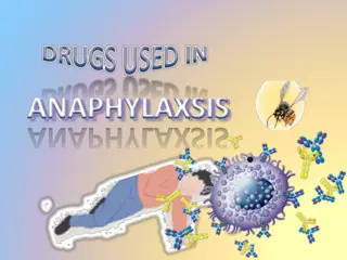
SHOCK AND ITS MANAGEMENT Dr. Santosh Y M Reader, Department of Shalyatantra
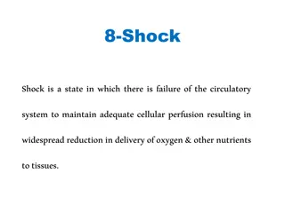
DEFINITION Shock is a state of poor perfusion with impaired cellular metabolism manifesting with severe pathophysiological abnormalities.
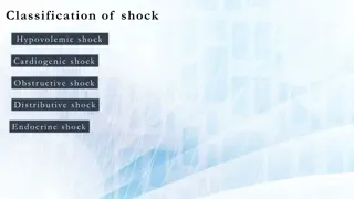
CLASSIFICATION HYPOVOLAEMIC SHOCK CARDIOGENIC SHOCK OBSTRUCTIVE DISTRIBUTIVE SEPTIC SHOCK NEUROGENIC SHOCK ANAPHYLATIC SHOCK
PATHOGENESIS In general, all forms of shock involve following 3 derangements: i) Reduced effective circulating blood volume. ii) Reduced supply of oxygen to the cells and tissues with resultant anoxia. iii) Inflammatory mediators and toxins released from shock induced cellular injury.
REDUCED EFFECTIVE CIRCULATING BLOOD VOLUME It may result by either of the following mechanisms: i) By actual loss of blood volume as occurs in hypovolaemic shock . ii) By decreased cardiac output without actual loss of blood (normovolaemia) as occurs in cardiogenic shock and septic shock.
IMPAIRED TISSUE OXYGENATION Reduction in the effective circulating blood volume from either of the above two mechanisms and from any of the etiologic agents, there is decreased venous return to the heart resulting in decreased cardiac output. This consequently causes reduced supply of oxygen to the organs and tissues and hence tissue anoxia occurs, which sets in cellular injury.
RELEASE OF INFLAMMATORY MEDIATORS In response to cellular injury, innate immunity of the body gets activated as a body defense mechanism and causes release of inflammatory mediators , but eventually these agents themselves become the cause of cell injury.
PATHOGENESIS OF CIRCULATORY SHOCK
RESPONSE OF INFLAMMATORY MEDIATORS IN SHOCK
HYPOVOLAEMIC SHOCK This form of shock results from inadequate circulatory blood volume by various etiologic factors that may be either from the loss of red cell mass and plasma due to haemorrhage or from the loss of plasma volume alone
CAUSES Acute haemorrhage . Dehydration from vomiting ,diarrhoea . Burn . Peritonitis, intestinal obstruction
PATHOGENESIS OF HYPOVOLAEMIC SHOCK Major effects in this are due to decreased cardiac output and low intra-cardiac pressure. Major clinical features are increased heart rate (tachycardia), low blood pressure (hypotension), low urinary output (oliguria to anuria) alteration in mental state (agitated to confused to lethargic).
CARDIOGENIC SHOCK Defined as circulatory failure with sudden fall in cardiac output caused due to acute diseases of heart without any actual reduction of blood(normovolaemia)
CAUSES a. Acute myocardial infarction, acute myocarditis b. Acute pulmonary embolism c. Drug induced d. Toxemia of any causes e. Cardiac conditions like valvular diseases, congenital heart diseases f. Cardiac compression: i. Cardiac tamponade due to collection of blood,pus, fluid in the pericardial space which prevents the heart to expand leading to shock. ii. Trauma to heart
PATHOGENESIS OF CARDIOGENIC SHOCK Dysfunction of RV- RV unable to pump blood to lungs, filling of left heart , as well as LV output. Dysfunction of LV- LV unable to maintain adequate stroke volume, LV output and systemic arterial blood pressure, pulmonary vasculature due to normal right ventricular output but failure of LV. engorgement of Compression shock- External compression decreases the cardiac output.
CLINICAL FEATURES Cold, clammy skin Hypotension Tachycardia Tachypnoea Oliguria Cyanosis Loss of consciousness
DIAGNOSIS Physical examination- assessment of vitals ECG Chest x-ray Investigations relevant to the cause
MANAGEMENT ACLS should be performed Proper oxygenation with intubation, ventilator support Avoiding fluid overload Antiarrythmic drugs Correction of electrolytes Rt sided failure due to pulmonary embolism- Large doses of heparin IV Left sided failure-If pain present morphine is given Diuretics in case of pulmonary edema Vasodilators, beta blockers, calcium channel blockers depending on the need Intra-aortic balloon pump(IABP) PTCA, CABG in severe conditions
SEPTIC SHOCK Shock induced due to severe bacterial infections or septicaemia. Causative agent- Gram +ve bacteria Gram ve bacteria Fungi Viruses Parasites
CAUSES Gram positive bacteria: Due to exotoxins Dissemination of potent exotoxin liberated into circulation. No evidence of bacteraemia No reduction in CO Hypotension will be there Urine output is normal Organisms- Clostridium tetani, Clostridium perfringens, Staphylococcus, streptococcus etc
CONT. Gram negative bacteria: Cause by endotoxins Most frequent source of septic shock Usual route is genitourinary system H/o urinary tract surgeries or instrumentation Second source- Respiratory system with h/o tracheostomy Third source- GIT with h/o biliary tract infections, intra-abdominal abscesses, peritonitis Common organism- E. coli, Klebsiella aerobacter, Proteus, Pseudomonas etc
PATHOGENESIS OF SEPTIC SHOCK
CLINICAL FEATURES Hyperdynamic shock (warm shock): Pyrogenic response is intact, so Development of chills Fever(>1000F) Hypotension Tachycardia Tachypnoea Increased cardiac output Reversible Diagnosis: Intermittent spikes of fever Bouts of chills
CONT. Hypodynamic hypovolaemic septic shock(cold septic shock): Pyrogenic response is lost,state of decompensated shock Features of hypovolaemia Decreased cardiac output Irreversible with MODS Anuria, respiratory failure, liver failure, cardiac depression, pulmonary edema, hypoxia Drowsiness>Coma>Death Diffcult to differentiate from hypovolaemic shock
MANAGEMENT Correction of fluid and electrolyte Culture & sensitivity test Appropriate antibiotics Treating of underlying cause Critical care, oxygen therapy, ventilator support Maintain blood pressure & urine output Short term high dose steroid therapy- Single dose of methylprednisolone or dexamethasone, may be repeated after 4 hours to avoid effects of endotoxaemia.
CRUSH SYNDROME It is a clinical condition caused by compression of muscle with subsequent rhabdomyolysis which can then lead to electrolyte imbalance, fluid sequestration & myoglobinuria. It is usually seen as a consequence of crush injuries. CAUSES- injuries, construction site accidents, bombings, use of torniquet for long period etc Earthquakes, landslides, mine
PATHOGENESIS Injury occurs Extravasation of blood into muscles at the site of injury Hypovolaemic shock Myohaemoglobin from crushed muscles enters systemic circulation Acute renal tubular necrosis Swelling of crushed muscles Compartment syndrome Circulation diminishes Ischaemic changes at affected site Severe pain, reduced UO, restlessness, delirium
MANAGEMENT Application of torniquet/pressure bandage to affected site as first aid Fluid resuscitation(upto 500ml) with monitoring of urine output Low molecular weight dextran(40000 units) Mannitol 1gm/kg IV 20% soln in 12 hours after catheterization Removal of torniquet Parallel incision to avoid compartment syndrome Blood transfusion only when renal function is normalised Haemodialysis in severe conditions
CONT 1. Activation of macrophage-monocytes Lysis of Gramnegative bacteria releases endotoxin, a lipopolysaccharide (LPS), into circulation where it binds to lipopolysaccharidebinding protein (LBP). The complex of LPS LBP binds to CD14 molecule on the surface of the monocyte/macrophages which are stimulated to elaborate proinflammatory cytokines, the most important ones being TNF and IL1.
By altering endothelial cell adhesiveness: This results in recruitment of more neutrophils which liberate free that cause vascular injury. b) Promoting nitric oxide synthase: This stimulates increased synthesis of nitric oxide which is responsible for vasodilatation and hypotension.
CONT.. II) ACTIVATION OF OTHER INFLAMMATORY RESPONSES Activation of mast cells: Histamine is released which increases capillary permeability. C) ACTIVATION OF COAGULATION SYSTEM: Enhances development of thrombi.
CONT D) ACTIVATION OF KININ SYSTEM: Released bradykinin causes vasodilatation and increased capillary permeability . Net result of above mechanisms is vasodilatation and increased vascular permeability in septic shock. Profound peripheral vasodilatation and pooling of blood causes hyperdynamic circulation in septic shock, in contrast to hypovolaemic and cardiogenic shock.
STAGES OF SHOCK 1. Compensated (nonprogressive, initial, reversible) shock . 2. Progressive decompensated shock . 3. Irreversible decompensated shock.
COMPENSATED (NON-PROGRESSIVE, INITIAL, REVERSIBLE) SHOCK WIDESPREAD VASOCONSTRICTION FLUID CONSERVATION BY THE KIDNEY STIMULATION OF ADRENAL MEDULLA
WIDESPREAD VASOCONSTRICTION In response to reduced blood flow (hypotension) and tissue anoxia, the neural and humoral factors (e.g. baroreceptors, chemo receptors,catecholamines, renin, and angiotensin II) are activated. All these bring about vasoconstriction, particularly in the vessels of the skin and abdominal viscera. Wide spread vasoconstriction is a protective mechanism as it causes increased peripheral resistance, increased heart rate (tachycardia) and increased blood pressure.
II) FLUID CONSERVATION BY THE KIDNEY In order to compensate the actual loss of blood volume in hypovolaemic shock, the following factors may assist in restoring the blood volume and improve venous return to the heart: a) Release of aldosterone from hypoxic kidney by activation of reninangiotens in aldosterone mechanism. b) Release of ADH due to decreased effective circulating blood volume. c) Reduced glomerular filtration rate (GFR) due to arteriolar constriction. d) Shifting of tissue fluids into the plasma due to lowered capillary hydrostatic pressure (hypotension).
III) STIMULATION OF ADRENAL MEDULLA In response to low cardiac output, adrenal medulla is stimulated to release excess of catecholamines (epinephrine and nonepinephrine) which increase heart rate and try to increase cardiac output.
CLASSICALFEATURESOF DECOMPENSATEDSHOCK i) Very low blood pressure ii) Subnormal temperature iii) Feeble and irregular pulse iv) Shallow and sighing respiration In addition, the patients in shock have pale face, sunken eyes, weakness, cold and clammy skin. .
NEUROGENICSHOCK due to sudden anxious or painful stimuli causing severe splanchnic vessel vasodilatation. Here, patient either goes for cardiac arrest and dies or recovers fully spontaneously
MORPHOLOGIC FEATURES OF SHOCK.
HYPOXIC ENCEPHALOPATHY If the blood pressure falls below 50 mmHg as occurs in systemic hypotension in prolonged shock and cardiac arrest, brain suffers from serious ischaemic damage with loss of cortical functions, coma, and a vegetative state.
HEARTINSHOCK Heart is affected in cardiogenic as well as in other forms of shock. There are 2 types of morphologic changes in heart in all types of shock: HAEMORRHAGES AND NECROSIS There may be small or large ischaemic areas or infarcts, particularly located in the sub epicardial and sub endocardial region.
SHOCKINLUNG Lungs due to dual blood supply are generally not affected by hypovolaemic shock but in septic shock the morphologic changes in lungs are quite prominent termed shock lung .
HAEMORRHAGIC GASTROENTEROPATHY The hypoperfusion of the alimentary tract in conditions such as shock and cardiac failure may result in mucosal and mural infarction called haemorrhagic gastroenteropathy . This type of nonocclusive ischaemic injury of bowel must be distinguished from fullfledged infarction in which deeper layers of the gut (muscularis and serosa) are also damaged. In shock due to burns, acute stress ulcers of the stomach or duodenum may occur and are known as Curling s ulcers.
COMPLICATIONS 1. Acute respiratory distress syndrome (ARDS) 2. Disseminated intravascular coagulation (DIC) 3. Acute renal failure (ARF) 4. Multiple organ dysfunction syndrome (MODS) With progression of the condition, the patient may develop stupor, coma and death
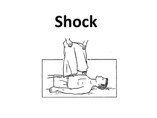
TREATMENT GENERAL MEASURES HOSPITALISATION . CARE OF ALL CRITICALLY ILL PATIENTS BEGIN WITH A, B And C. OXYGEN SHOULD BE ADMINISTERED BY FACE MASK TO ALL PATIENTS WHO ARE CONSCIOUS AND ARE ABLE TO MAINTAIN THEIR AIRWAY .
CONT If unconscious endotracheal intubation and ventilation with oxygen may be necessary . Haemorrhage control . Intravenous access : urgent administration of ringers lactate to restore blood volume to normal . Investigations : blood collected for routine investigations as well as for blood grouping and cross matching
CONT. PRESSURE & PACKING POSITION & REST Sedation to relieve anxiety midazolam in titrated doses of 1-2mg intravenously may be given .


 FLUID CONSERVATION")
 STIMULATION OF ADRENAL")
 STIMULATION OF ADRENAL")