Understanding the Anatomy of the Trachea
Explore the detailed structure and functions of the trachea, including its position, associated structures, composition, and blood/nerve supply. Learn about the relationship of the trachea with surrounding organs and its essential role in the respiratory system.

Download Presentation

Please find below an Image/Link to download the presentation.
The content on the website is provided AS IS for your information and personal use only. It may not be sold, licensed, or shared on other websites without obtaining consent from the author. If you encounter any issues during the download, it is possible that the publisher has removed the file from their server.
You are allowed to download the files provided on this website for personal or commercial use, subject to the condition that they are used lawfully. All files are the property of their respective owners.
The content on the website is provided AS IS for your information and personal use only. It may not be sold, licensed, or shared on other websites without obtaining consent from the author.
E N D
Presentation Transcript
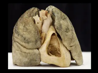
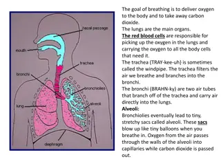
Position The trachea or windpipe is a continuation of the larynx and extends downwards to about the level of the 5ththoracic vertebra where it divides (bifurcates) at the carina into the right and left bronchi, one bronchus going to each lung. It is approximately 10 to 11 cm long and lies mainly in the median plane in front of the oesophagus
Structures associated with the trachea Superiorly the larynx Inferiorly the right and left bronchi Anteriorly upper part: the isthmus of the thyroid gland lower part: the arch of the aorta and the sternum Posteriorly the oesophagus separates the trachea from the vertebral column Laterally the lungs and the lobes of the thyroid gland.
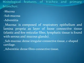
Structure The trachea is composed of from 16 to 20 incomplete (C-shaped) rings of hyaline cartilages lying one above the other. The cartilages are incomplete posteriorly. Connective tissue and involuntary muscle join the cartilages and form the posterior wall where they are incomplete. The soft tissue posterior wall is in contact with the oesophagus . There are three layers of tissue which 'clothe' the cartilages of the trachea. The outer layer. This consists of fibrous and elastic tissue and encloses the cartilages.
The relationship of the trachea to the oesophagus.
The middle layer. This consists of cartilages and bands of smooth muscle that wind round the trachea in a helical arrangement. There is some areolar tissue, containing blood and lymph vessels and autonomic nerves. The inner lining. This consists of ciliated columnar epithelium, secreting goblet cells. containing mucus-
Blood and nerve supply, lymph drainage The arterial blood supply. This is mainly by the inferior thyroid and bronchial arteries and the venous return is by the inferior thyroid veins into the brachiocephalic veins. The nerve supply. This is by parasympathetic and sympathetic fibres. Parasympathetic supply is by the recurrent laryngeal nerves and other branches of the vagi. Sympathetic supply is by nerves from the sympathetic ganglia.
The trachea and some of its associated structures.
Functions Support and patency. Mucociliary escalator. Cough reflex. Warming, humidifying and filtering of air
BRONCHI AND SMALLER AIR PASSAGES The two primary bronchi are formed when the trachea divides, i.e. about the level of the 5th thoracic vertebra. The right bronchus. This is wider, shorter and more vertical than the left bronchus and is therefore the more likely of the two to become obstructed by an inhaled foreign body. It is approximately 2.5 cm long. After entering the right lung at the hilum it divides into three
branches, one to each lobe. Each branch then subdivides into numerous smaller branches. The left bronchus. This is about 5 cm long and is narrower than the right. After entering the lung at the hilum it divides into two branches, one to each lobe. Each branch then subdivides into progressively smaller tubes within the lung substance.
Bronchi and bronchioles Structure The bronchi are composed of the same tissues as the trachea. They are lined with ciliated columnar epithelium. The bronchi progressively subdivide into bronchioles terminal bronchioles, respiratory bronchioles, alveolar ducts and finally, alveoli. Towards the distal end of the bronchi the cartilages become irregular in shape and are absent at bronchiolar level.
Blood and nerve supply, lymph drainage right and left bronchial arteries and the venous return is mainly through the bronchial veins The nerve supply. This is by parasympathetic and sympathetic nerves. The vagus nerves (parasympathetic) stimulate contraction of smooth muscle in the bronchial tree, causing bronchoconstriction, and sympathetic stimulation causes bronchodilatation
The lymphatic vessels and lymph nodes. Lymph is drained from the walls of the air passages in a network of lymph vessels. It passes through lymph nodes situated around the trachea and bronchial tree then into the thoracic duct on the left side and right lymphatic duct on the other.
Functions of air passages not involved in gaseous exchange Control of air entry. The following functions continue as in the upper airways: warming and humidifying support and patency removal of particulate matter cough reflex.
Respiratory bronchioles and alveoli Lobules are the blind ends of the respiratory tract distal to the terminal bronchioles, consisting of: respiratory bronchioles, alveolar ducts and alveoli (tiny air sacs) It is in these structures that the process of ga exchange occurs. The walls gradually become thinner until muscle and connective tissue fade out leaving a single layer of simple squamous epithelial cells in the alveolar ducts and alveoli.
The alveoli are surrounded by a network of capillaries. The exchange of gases during respiration takes place across two membranes, the alveolar and capillary membranes.
Interspersed between the squamous cells are other cells that secrete surfactant, a phospholipid fluid which prevents the alveoli from drying out. In addition, surfactant reduces surface tension and prevents alveolar walls collapsing during expiration. Secretion of surfactant into the distal air passages and alveoli begins about the 35th week of fetal life. Its presence in newborn babies facilitates expansion of the lungs and the establishment of respiration. It may not be present in sufficient amounts in the immature lungs of premature babies, causing difficulty in establishing respiration.