Understanding the Cardiovascular System and Blood Composition
Explore the intricate workings of the cardiovascular system and the composition of blood. Learn about the components, functions, and importance of blood circulation in the body. Discover how the heart, blood vessels, and various blood cells work together to maintain overall health and well-being.

Download Presentation

Please find below an Image/Link to download the presentation.
The content on the website is provided AS IS for your information and personal use only. It may not be sold, licensed, or shared on other websites without obtaining consent from the author. If you encounter any issues during the download, it is possible that the publisher has removed the file from their server.
You are allowed to download the files provided on this website for personal or commercial use, subject to the condition that they are used lawfully. All files are the property of their respective owners.
The content on the website is provided AS IS for your information and personal use only. It may not be sold, licensed, or shared on other websites without obtaining consent from the author.
E N D
Presentation Transcript
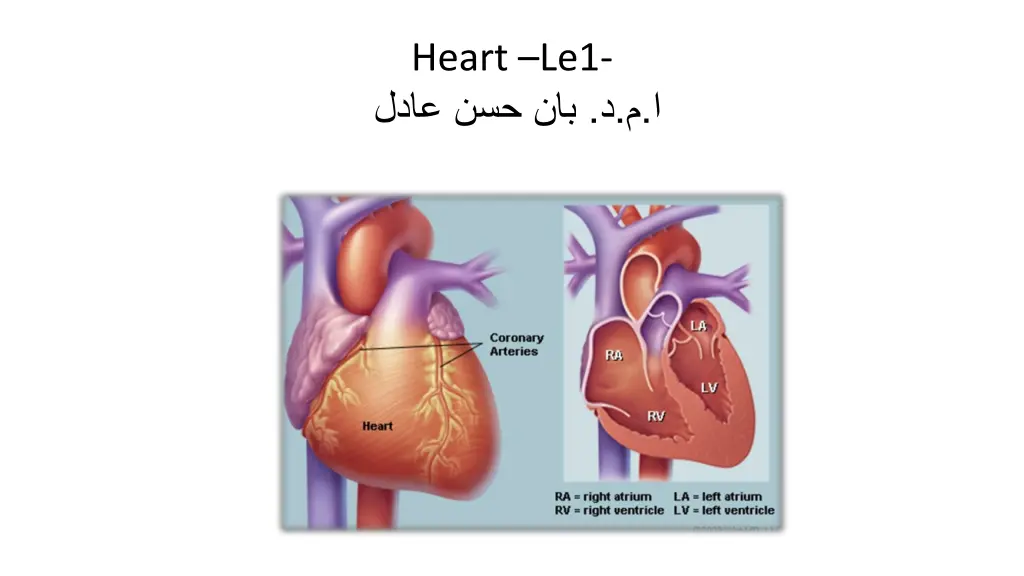
Heart Le1- . . .
Physics of the Cardiovascular System (Cardiovascular system (CVS): blood, vessels, heart) Cardiovascular system blood heart vessels
The cells of the body act like individual engines. In order for them to function they must have: Regulation Transportation hormonal protection temperatur immune respiratory nutritive excretory e Carry hormones to O2,CO2 Carry absorbed digestion products to liver and to tissue against pathogens Carry metabolic wastes kidneys be excreted Blood clotting Complement act Divert blood to cool or warm target tissue to to the body
Component of circulatory system Blood vessels Heart Lymphatic system veins Arteries capillaries
The blood and its supply of O2 are so important to the body that the heart is the first major to develop in the embryo. * Fetal heart 1. Start blood circulation at the eighth week after conception 2. Obtain oxygenated blood from mother via umbilical cord 3. An opening between RA and LA 90% of blood flows from RA to LA through the opening 10% circulates through fetal lungs Within minutes after birth, the opening is closed in effect Complete closure takes several months Blue baby: inadequate closure of the opening, requires a surgery
The blood 1. The red color is caused by red blood cells (erythrocytes), flat disks about 7 m in diameter, which is represent 45% of the volume of blood. 2. White blood cells (leukocytes), present in small amounts. White blood cells ( 9 to 15 m in diameter), play an important role in combating disease. There are about 8000 white blood cells/mm3 of blood. When there is an infection in the body the number of white blood cells (white count) increase. (In one type of blood cancer, leukemia, there is an excessive production of white blood cells). 3. Platelets ( 1 to 4 m in diameter) are involved in the clotting function of blood. There are about 3 103 platelets/mm3 of blood. 4. Plasma is clear fluid, accounts for the other 55%. The combination of red blood cells and plasma causes blood to have flow properties different from those of a fluid like water.
The blood erythrocytes Plasma leukocytes Platelets
Major components of the cardiovascular system l. Pulmonary circulation 2. Systemic circulation in the rest of the body The blood is pumped by the contraction of the heart muscles from the left ventricle at a pressure of about 125mmHg into a system of arteries that Subdivided into smaller arteries (arterioles) and to capillary bed. During the a few seconds it is in the capillary bed the blood supplies O2 to the cells and picks up CO2 from the cells. After passing through the capillary bed the blood collects in small veins that gradually combine into larger veins before inters the right side of the heart. The returning blood is momentarily stored in the reservoir (the right atrium), and during a weak contraction (5 to 6mmHg) the blood flows into right ventricle. On the next ventricular contraction this blood is pumped at a pressure of about 25mmHg. Via the pulmonary arteries to the capillary system in the lungs, where it receives more O2 and where some of the CO2 diffuses into the air in the lungs to be exhaled. The freshly oxygenated left reservoir of the heart (left atrium): during weak contraction (7 to 8mmHg), the blood flows into the left ventricle. On the next ventricular contraction this blood is again pumped from the left side of the heart into the general circulation.
How fast dose your blood flow The blood goes from the aorta into the smaller arteries and arterioles with greater total-cross sectional areas the velocity of the blood decreases: The velocity= flow rate/ cross sectional The average velocity in the aorta 30cm/s; that in a capillary is only about 1mm/s, this low velocity allows time for diffusion of gases to occur The flow rate due to Poiseuilles law = P ( /8) (1/?) (R4/L)
The factors affect the flows of blood in the vessel are. 1. Viscosity (?), the cgs unit used to measure viscosity is Poise, the SI unit for viscosity is Pascal second (Pas), which equals 10 poises. The viscosity of blood is typically 3 10-3 to 4 10-4 pas. 2. Pressure difference ( P). If P is doubled, the flow rate also doubles. 3. The length (L), the flow varies inversely with the length 4. The radius (R), if the radius is doubled the flow rate increases by 24 or factor 16.
Work done by the heart Each contraction of the heart muscles forces about 80ml of blood through the lungs from the right ventricle and a similar volume from the left ventricle. In the process the heart does work. The pressure in two pumps of the heart are not the same. In the pulmonary system the pressure is low because of low resistance of the blood vessels in the lungs. The maximum pressure (systole) about 25mmHg. In order to circulate the blood through the much larger systemic net work the left side of the heart muscle produce pressure about 120mmHg at the peak (systole) of each cardiac cycle. The work W done by a pump working at a constant pressure P is equal to the product of pressure and volume pumped V or W = P V Average P = 10cmHg V = 80ml W= pgh V = 13.6 g/cm3 980 cm/s2 10cm 80cm3 = 1.1 107 ergs
Cardiac Contraction As mentioned above, a series of events occur in a specific order during a normal heartbeat. This process is called the cardiac cycle. The cardiac cycle can be broken down into two components, systole- and diastole. Diastole occurs when the heart muscle is relaxed and begins to fill with venous blood_ in the right atrium and oxygenated blood in the left atrium. Systole is the time when the heart contracts. During systole, the heart forces oxygenated blood out of the left ventricle and deoxygenated blood to the lungs through the right ventricle.
The human heart contains four valves: tricuspid valve, pulmonic valve, mitral valve and aortic valve.
During the first diastole period, the atria and ventricles are relaxed and the atrioventricular valves are open. Oxygen-depleted blood returning to the heart from the body passes through the superior and inferior vena cavae and flows to the right atrium. The open atrioventricular valves (tricuspid and mitral valves) allow blood to pass through the atria to the ventricles. Impulses from the sinoatrial (SA) node atrioventricular (AV) node and the AV node send signals that trigger both atria to contract. As a result of the contraction, the right atrium empties its contents into the right ventricle. The tricuspid valve, located between the right atrium and right ventricle, prevents blood from flowing back into the right atrium. travel to the
At the beginning of the first systole period, the right ventricle is filled with blood passed on from the right atrium. The ventricles receive impulses from fiber branches (Purkinje fibers), which carry electrical impulses to the ventricles causing them to contract. As this occurs, the atrioventricular valves close and the semilunar valves (pulmonary and aortic valves) open. Ventricular contraction causes oxygen-depleted blood from the right ventricle to be pumped to the pulmonary artery. The pulmonary valve prevents blood from flowing back into the right ventricle. The pulmonary artery carries oxygen- depleted blood along the pulmonary circuit to the lungs. There, blood picks up oxygen and is returned to the left atrium of the heart by the pulmonary veins.
In the second diastole period, the semilunar valves close and the atrioventricular valves open. Oxygenated blood from the pulmonary veins fills the left atrium (blood from the venae cavae is also filling the right atrium at this time). The SA node contracts again triggering both atria to contract. Atrial contraction causes the left atrium to empty its contents into the left ventricle (the right atrium is also emptying blood into the right ventricle at this time). The mitral valve, located between the left atrium and left ventricle, prevents oxygenated blood from flowing back into the left atrium.
During the second systole period, the atrioventricular valves close and the semilunar valves open. The ventricles receive impulses and contract. Oxygenated blood in the left ventricle is pumped to the aorta and the aortic valve prevents the oxygenated blood from flowing back into the left ventricle (oxygen-depleted blood is also being pumped from the right ventricle to the pulmonary artery at this time). The aorta branches out to provide oxygenated blood to all parts of the body through systemic circulation. After its tour through the body, oxygen-depleted blood is returned to the heart via the venae cavae.
The physics of some cardiovascular diseases Heart diseases often have physical component Many of these diseases, for example, increase the work load of the heart or reduce its ability to work at a normal rate. W = P V equation of work done The work done by the heart is roughly the tension of the heart muscle times how long it acts. Anything that increase the work load of the heart. For example: 1-Hypertension Causes the muscle tension to increase in proportion to the pressure, due to Laplace law T = PR so the high blood pressure causes to increase the work done by equation W = P V. 2- Tachycardia A fast heart rate increases the work load since the amount of time the heart muscle spend contracting increases. 3- Enlargement of the heart and reduction in the ability of the heart to provide adequate circulation. If the radius of the heart is doubled, the tension of the heart muscle must also be doubled if the same pressure is to maintained. Since the heart muscle is stretched, it may not be able to produce sufficient force to maintain normal circulation; the stretched heart muscle is also much less efficient than normal heart muscle.
Electrical signal from the heart- the electrocardiogram Introduction The body relies on the heart to circulate blood throughout the body. The heart is responsible for pumping oxygenated blood from the lungs out to the body through the arteries and also circulating deoxygenated blood back to the lungs. from the body through the veins. The heart is divided into four chambers and each chamber is responsible for a different part of the circulatory process mentioned above. Deoxygenated blood first enters the right atrium via the vena cava, where it is then pumped into the right ventricle. The right ventricle pumps this deoxygenated blood through the pulmonary artery to the lungs, where it flows through the alveoli, receives oxygen, and then is returned to the heart through the pulmonary vein and into the left atrium. The left atrium then pumps this oxygenated blood into the left ventricle, where then it is pumped out to the rest of the body through the aorta. This process of contracting the different chambers is highly coordinated and the coordination is controlled by specialized regions of the heart responsible for electrical stimulation of cardiac muscle.
Life several other bioelectrical signals, the electrical impulses generated by the heart can be measured on the surface of the skin with electrodes. Using surface electrodes, the cardiac potential of the heart can be measured and correlated with regions of cardiac excitation. This measurement is called an electrocardiogram (ECG). The ECG can be used to evaluate cardiac function, heart rate, and cardiac arrhythmias. The electrical activation that creates the normal heartbeat can in some instances cause abnormal cardiac function. Disorders such as bradycardia (slow heart rate), tachycardia (fast heart rate)and electrical conduction problems such as bundle branch blocks can be all diagnosed from the ECG.


