Exploring the anatomy of the eye ball, including its three coats (fibrous, vascular pigmented, nervous) and components like the sclera, cornea, choroid, ciliary body, and iris. Learn about the structure and function of each part in detail.
Please find below an Image/Link to download the presentation.
The content on the website is provided AS IS for your information and personal use only. It may not be sold, licensed, or shared on other websites without obtaining consent from the author. If you encounter any issues during the download, it is possible that the publisher has removed the file from their server.
You are allowed to download the files provided on this website for personal or commercial use, subject to the condition that they are used lawfully. All files are the property of their respective owners.
The content on the website is provided AS IS for your information and personal use only. It may not be sold, licensed, or shared on other websites without obtaining consent from the author.
E N D
Presentation Transcript
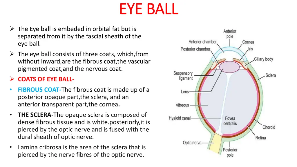
EYE BALL EYE BALL The Eye ball is embeded in orbital fat but is separated from it by the fascial sheath of the eye ball. The eye ball consists of three coats, which,from without inward,are the fibrous coat,the vascular pigmented coat,and the nervous coat. COATS OF EYE BALL- FIBROUS COAT-The fibrous coat is made up of a posterior opaque part,the sclera, and an anterior transparent part,the cornea. THE SCLERA-The opaque sclera is composed of dense fibrous tissue and is white.posteriorly,it is pierced by the optic nerve and is fused with the dural sheath of optic nerve. Lamina cribrosa is the area of the sclera that is pierced by the nerve fibres of the optic nerve.
FIBROUS COAT CONT. FIBROUS COAT CONT. The sclera is also pierced by the ciliary arteries and nerves and their associated veins,the venae vorticosae. The sclera is directly continuous infront with the cornea at the corneoscleral junction,or limbus. THE CORNEA-The transparent cornea is largely responsible for the refraction of the light entering the eye. It is in contact posteriorly with the aqueous humor. Cornea is avascular&devoid of lymphatic drainage ,it is nourished by diffusion from the aqueous humor. supplied by long ciliary nerve from the ophthalmic division of trigeminal nerve. The cornea is the most important refractive medium of the eye.
VASCULAR PIGMENTED COAT VASCULAR PIGMENTED COAT The vascular pigmented coat consists ,from behind forward,of the choroid the ciliary body,and the iris. The choroid is composed of an outer pigmented layer and an inner, highly vascular layer. THE CILIARY BODY-The ciliary body is continuous posteriorly with the choroid,and anteriorly it lies behind the peripheral margin of the iris.it is composed of the ciliary ring,the ciliary process and the ciliary muscle. CILIARY RING-is the posterior part of the body,and its surface has shallow grooves,the ciliary striae. CILIARY PROCESS-are radially arranged folds,or ridges,to the posterior surfaces of which are connected the suspensory ligaments of the lens.
CILIARY MUSCLES CILIARY MUSCLES Is composed of meridinal and circular fibres of smooth muscles. The meridinal fibres run backward from the region of the corneoscleral junction to the ciliary processes.the circular fibres are fewer in number and lie internal to the meridinal fibres. NERVE SUPPLY-The ciliary muscle is supplied by the parasympathetic fibres from the oculomotor nerve. After synapsing in the ciliary ganglion,the postganglionic fibres pass forward to the eye wall in the short ciliary nerves. ACTION-Contraction of ciliary muscle,especially the meridinal fibres,pulls the ciliary fibres forward.this relieves the tension in the suspensory ligament,and the elastic lens becomes more convex.this increases the refractive power of the lens.
THE IRIS & PUPIL The iris is a thin, contractile,pigmented diaphragm with a central aperture,the pupil.it is suspended in the aqueous humor between the cornea and the lens. The periphery of the iris is attached to the ant surface of the ciliary body. It divides the space between the lens and the cornea in to an ant &post chamber. The muscle fibres of the iris are involuntary & consists of circular & radial fibres. The circular fibres form the sphincter pupillae & are arranged around the margin of the pupil. The radial fibres form the dilator pupillae& consist of a thin sheet of radial fibres that lie close to the posterior surface.
NERVE SUPPLY- The sphincter pupillae is supplied by parasymphathetic fibres from the oculomotor nerve.after synapsing in the ciliary ganglion,the postganglionic fibres pass forward to the eye ball in the short ciliary nerves.the dilator pupillae is supplied by the sympathetic fibres,which pass forward to eye ball in the long ciliary nerves. ACTION-The sphincter pupillae constricts the pupil in the presence of bright light and during accomodation.the dilator pupillae dilates the pupil in the presence of light of low intensity or in the presence of excessive sympathetic activity such as occurs in fright.
NERVOUS COAT:THE RETINA NERVOUS COAT:THE RETINA The retina consists of an outer pigmented layer and an inner nervous layer. Its outer surface is in contact with the choroid,and its inner surface is in contact with the vitreous body. The post three fourths of the retina is the receptor organ. Its ant edge forms a wavy ring the orra serrata and the nervous tissues end here. The ant part of the retina is nonreceptive and consists of pigment cells,with a deeper layer of columnar epithelium. This ant part of the retina covers the ciliary processes and the back of iris.
RETINA CONT. At the center of the post part of retina is an oval yellowish area,the macula lutea,which is the area of the retina for the most distinct vision. It has a central depression the fovea centralis. The optic nerve leaves the retina about 3 mm to the medial side of macula lutea by the optic disc. The optic disc is slightly depressed at its center where it is pierced by the central artery of the retina at the optic disc. At the optic disc is a complete absence of rods and cones so that it is insenstive to light and is referred to as the blind spot. On ophthalmological examination,the optic disc is seen to be pale pink in colour,much paler than the surrounding retina.
CONTENTS OF THE EYE BALL CONTENTS OF THE EYE BALL The contents of the eye ball consist of the refractive media,the aqueous humor,the vitreous body,and the lens. AQUEOUS HUMOR-The aqueous humor is a clear fluid that fills the ant & post chambers of the eye ball. It is believed to be a secretion from the ciliary process,from which it enters the post chamber. It then flows in to the ant chamber through the pupil and is drained away through the spaces at the irido corneal angle in to the canal of schlemm. Obstruction to the draining of the aqueous humor results in a rise in intraocular pressure called glaucoma.this can produce degenerative changes in the retina with consequent blindness. The function of the aqueous humor is to support the wall of the eye ball by exerting internal pressure and thus maintaining optical shape. It also nourishes cornea & lens& removes the products of metabolism.
VITREOUS BODY VITREOUS BODY The vitreous body fills the eye ball behind the lens and is transparent gel. The hyaloid canal is a narrow channel that runs through the vitreous body from the optic disc to the post surface of the lens; in the fetus,it is filled by the hyaloid artery,which disappears before birth. The function of the vitreous body is to contribute slightly to the magnifying power of the eye. It supports the post surface of the lens and assists in holding the neural part of the retina against the pigmented part of the retina.
THE LENS THE LENS The lens is a transparent ,biconvex structure,enclosed in a transparent capsule. It is situated behind the iris and infront of the vitreous body and is encircled by ciliary processes. The lens consists of an elastic capsule,which envelops the structure;a cuboidal epithelium,which is confined to the ant surface of the lens;and lens fibres which are formed from the cuboidal epithelium at the equator of the lens.the lens fibres make up the bulk of the lens. The equatorial region or circumference of the lens is attached to the ciliary processes of the ciliary body by the suspensory ligament.the pull of the radiating fibres of the suspensory ligament tends to keep the elastic lens flattened so that the eye can be focussed on distant object.