Understanding the Cross-Bridge Cycle in Muscle Contraction
Learn about the cross-bridge cycle in muscle contraction, where actin and myosin interact to drive the sliding of filaments. Explore how ATP, calcium ions, and conformational changes play crucial roles in this process. Understand the stages of binding, power generation, and unbinding that enable muscle movement.

Download Presentation

Please find below an Image/Link to download the presentation.
The content on the website is provided AS IS for your information and personal use only. It may not be sold, licensed, or shared on other websites without obtaining consent from the author. If you encounter any issues during the download, it is possible that the publisher has removed the file from their server.
You are allowed to download the files provided on this website for personal or commercial use, subject to the condition that they are used lawfully. All files are the property of their respective owners.
The content on the website is provided AS IS for your information and personal use only. It may not be sold, licensed, or shared on other websites without obtaining consent from the author.
E N D
Presentation Transcript
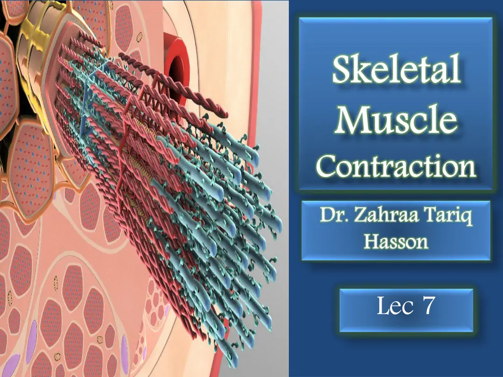
The The Cross Cross- -bridge bridge Cycle Cycle During muscle contraction, the mechanism that drives the sliding of thick and thin filaments past one another is called the cross-bridge cycle. Actin filament is composed of double stranded F-actin protein that wrap around each other as a helix. Tropomyosin protein blocks attachment sites of myosin on actin molecule and thus prevent cross-bridge cycle while troponin attaches to tropomyosin.
Cross Bridge cycle: Binding of actin and myocin Before contraction begins, the heads of the cross- bridges bind with ATP. The ATPase activity of the myosin head immediately cleaves the ATP but leaves the cleavage products, ADP plus phosphate ion, bound to the head. In this state, the conformation of the head is such that it extends perpendicularly toward the actin filament but is not yet attached to the actin.
Power When the troponin-tropomyosin complex binds with calcium ions, active sites on the actin filament are uncovered, and the myosin heads then bind with these . The active change arm This filament energy occurred earlier Power stroke stroke: : he bond active site change in in the arm of of the This provides filament. . The energy already occurred in in the earlier. . bond between site of of the the head, the cross provides the The energy already stored, the head between the the actin head, prompting cross- -bridge the power energy that stored, by head when the head actin filament prompting the bridge. . power stroke that activates by the when the head of of the filament causes the head the cross causes a a conformational head to to tilt cross- -bridge conformational tilt toward bridge and and the the toward the the stroke for activates the the conformational the ATP for pulling the power conformational change ATP molecule pulling the power stroke the actin stroke is is the change that was cleaved actin the that cleaved molecule was
Unbinding A new ATP enters the ATPase site on the myosin head, triggering a conformational change in the head, which decreases the affinity of myosin for actin, so the myosin detaches from the actin. Soon after it binds to myosin s ATPase site, ATP is split by hydrolysis into ADP and Pi, which releases energy. Some of the energy is captured by the myosin molecule as it goes into its high-energy conformation. Although ATP has been hydrolyzed at this point, the end- products of the reaction (ADP and Pi) remain bound to the ATPase site. If calcium is present, the cycle will continue by revisiting step 1. Unbinding of of Myosin Myosin and and Actin Actin: :
At any given time, some cross-bridges are starting the cycle, others are finishing it, and still others are at various stages in between and this help maintain muscular contraction. When the cross-bridge cycle stops and the contraction ends, the thin filaments passively slide back to their original position. A skeletal muscle fiber is capable of shortening to approximately 60% of its resting length. The cross- bridge cycle could continue indefinitely, so long as there is abundant ATP. To prevent this never-ending cycle from happening, the regulatory proteins troponin and tropomyosin interact with calcium, controlling the availability of myosin-binding sites on actin and thereby regulating the cross-bridge cycle.
Skeletal Muscle Tone. Even when muscles are at rest, a certain amount of tautness usually remains, which is called muscle tone. Because normal skeletal muscle fibers do not contract without an action potential to stimulate the fibers, skeletal muscle tone results entirely from a low rate of nerve impulses coming from the spinal cord.
Muscle Fatigue: Muscle fatigue occurs when a muscle can no longer sustain its contraction during prolonged or intense physical activity. Causes of of Muscle The primary cause is the depletion of muscle glycogen, which serves as the primary energy source for muscle fibers. As glycogen stores are exhausted, the muscle's ability to generate ATP diminishes, leading to a reduction in contractile force. Causes Muscle Fatigue Fatigue 1.
Another neuromuscular acetylcholine release and receptor activation become less efficient after extended activity, impairing muscle contraction. contributing factor is the reduced where 2. junction transmission, Additionally, the interruption of muscle perfusion (blood flow) leads to rapid fatigue (within 1-2 minutes) due to the loss of oxygen and nutrients necessary for cellular energy production. 3.
Myasthenia Gravis Myasthenia gravis is a serious autoimmune disorder characterized by muscle weakness and fatigue, often triggered by repetitive activity. It primarily affects women in their 20s and men in their 60s. The disease occurs due to antibodies targeting nicotinic acetylcholine neuromuscular transmission. There are two main forms: one affecting extraocular muscles, and the other causing generalized skeletal muscle weakness. Symptoms improve with rest or cholinesterase inhibitors(drugs), which breakdown, enhancing neurotransmitter availability. receptors, impairing block acetylcholine
Rigor Mortis: Several hours after death, all muscles enter a state of contracture called "rigor mortis," where muscles contract and become stiff without action potentials. This rigidity occurs due to the depletion of ATP, which is required to detach cross-bridges from actin filaments during muscle relaxation. Muscles remain in rigor until muscle proteins degrade, typically 15 to 25 hours later, due to autolysis from lysosomal enzymes. These processes occur more rapidly at higher temperatures.
Muscle Muscle hypertrophy refers to an increase in muscle mass, while muscle atrophy refers to its decrease. Hypertrophy results from an increase in actin and myosin filaments within muscle fibers, leading to fiber hypertrophy. This process is amplified by muscle loading during contraction, with significant hypertrophy occurring after just a few strong contractions daily over 6 to 10 weeks. Muscle atrophy is the reduction in muscle mass and strength, often due to disuse, aging, or certain medical conditions. Muscle Hypertrophy Hypertrophy and and Muscle Muscle Atrophy Atrophy: :
It occurs when there is a decrease in the size of muscle fibers, primarily due to a reduction in the synthesis of proteins like actin and myosin. This process is driven by an imbalance between protein degradation and protein synthesis, with the former dominating in atrophic muscles